In multicellular organisms, apoptosis, commonly referred to as programmed cell death, is a tightly controlled process that is crucial for tissue homeostasis, development, and the removal of unhealthy or unneeded cells. This article provides an in-depth analysis of apoptosis, outlining its definition, routes, test techniques, applications, and contrast with necrosis.
Table of Contents
Definition of Apoptosis
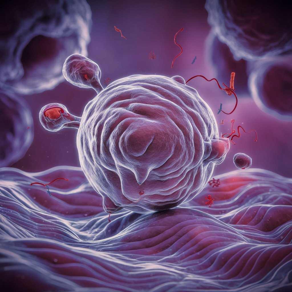
One kind of cell death known as apoptosis is marked by certain morphological and biochemical changes, such as membrane blebbing, chromatin condensation, DNA fragmentation, shrinking of the cell, and the creation of apoptotic bodies. These alterations are the result of a series of biological actions regulated by certain genes and signaling pathways.
Pathways of Apoptosis
1. Intrinsic Pathway (Mitochondrial Pathway)
Causes mitochondrial outer membrane permeabilization (MOMP) when it is activated by intracellular stressors such DNA damage, oxidative stress, or growth factor depletion. This triggers the caspase cascade by activating caspase-9 and releasing cytochrome c into the cytoplasm.
2. Extrinsic Pathway (Death Receptor Pathway)
started when outside signals attach to death receptors (such as when a ligand binds to a Fas receptor), creating the death-inducing signaling complex (DISC). This initiates caspase-8, which can then either directly activate executioner caspases or, by Bid cleavage, engage the intrinsic route.
Assay Methods
1. Morphological Assays
These include the TUNEL assay for DNA fragmentation, Annexin V staining for phosphatidylserine externalization, and staining methods like Hoechst or DAPI for chromatin condensation.
2. Biochemical Assays
Utilizing fluorogenic substrates, Western blotting for cleaved caspases, PARP cleavage, and DNA ladder assays for DNA fragmentation, one may quantify caspase activity.
3. Flow Cytometry
uses Annexin V/PI staining to identify apoptosis and differentiate between late apoptotic/necrotic (Annexin V positive, PI positive) and early apoptotic (Annexin V positive, PI negative) cells.
4. Live-Cell Imaging
employing fluorescent dyes or genetically encoded reporters for it markers to monitor apoptotic events in real time.
Examples
1. Embryonic Development
During embryogenesis, apoptosis is essential for shaping tissues and organs because it gets rid of undesirable cells or structures.
2. Immune System Regulation
By eradicating autoreactive or activated lymphocytes via processes including activation-induced cell death (AICD), it controls immune cell populations.
3. Tissue Homeostasis
Apoptosis eliminates aging or damaged cells from adult tissues to preserve homeostasis and avoid accumulation and possible tumor development.
4. Pathological Conditions
Numerous illnesses, such as cancer, autoimmune diseases, and neurodegenerative conditions like Alzheimer’s disease, are influenced by dysregulated apoptosis.
Comparison with Necrosis
Apoptosis
unique morphological and molecular characteristics associated with controlled, predetermined cell death. It doesn’t cause inflammation and helps nearby phagocytes remove cells more easily.
Necrosis
Uncontrolled, accidental cell death typically resulting from physical or chemical damage. It leads to cell swelling, rupture, and inflammation, eliciting an immune response. Necrotic cells release damage-associated molecular patterns (DAMPs), activating inflammatory pathways.
Frequently Asked Question
1. What are the assays for necrosis?
The two main types of cytotoxicity assays used to quantify necrosis are those based on the differential uptake of DNA binding dyes (like propidium iodide) that do not cross the plasma membrane of living cells and those based on the leakage of intracellular molecules through.
2. What is apoptosis and give an example?
Definition: 0:00. The process of planned cell death is called apoptosis. It is used in the early stages of development to get rid of undesirable cells, such the ones that grow between a developing hand’s fingers. Adults employ to get rid of cells that are too damaged to be repaired.
3. What is the apoptosis assay?
The biological processes involved in programmed cell death, such as caspase activation, phosphatidylserine (PS) exposure on the cell surface, and DNA fragmentation, are identified and measured using an that test.
4. What is necrosis vs apoptosis assay kit?
This kit can identify necrosis and they-related cell death simultaneously. It may be applied to evaluate how new medicinal drugs affect the integrity of cells. Use flow cytometry or fluorescence microscopy to analyze the fluorescent signal.
Related Article