Acridine Orange Staining: Simple Explanation

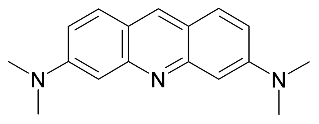
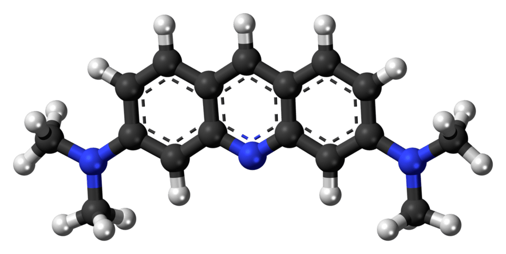
Acridine orange staining is a technique used in laboratories to detect and differentiate between DNA and RNA in cells. It is especially useful for identifying certain infections, assessing cell health, and diagnosing conditions like cancer. This method uses a special fluorescent dye called acridine orange, which binds to genetic material in cells (DNA and RNA) and makes it glow under specific lighting conditions, like ultraviolet (UV) light.
Table of Contents
Here’s a breakdown of everything you need to know about acridine orange staining, in simple terms:

Principle of Acridine Orange Staining
Acridine orange is a dye that interacts with the genetic material (DNA and RNA) inside cells. The way it interacts with DNA and RNA depends on the structure of these molecules. When acridine orange binds to double-stranded DNA (dsDNA), it emits a green fluorescence under UV light. When it binds to single-stranded DNA (ssDNA) or RNA, it emits a red or orange fluorescence.
This difference in the color of fluorescence allows scientists to distinguish between DNA and RNA in cells. Essentially, acridine orange helps scientists “see” these molecules by making them glow, which can be particularly helpful in medical diagnoses, cell health assessment, and research.
Requirements for Acridine Orange Staining
Before performing acridine orange staining, you need the following items:
- Acridine Orange Dye: This is the main reagent that binds to DNA and RNA.
- Phosphate-buffered saline (PBS): A buffer solution used to maintain the cells in an environment similar to their natural state.
- Sample: This could be a smear of cells from tissues, a blood sample, or a bacterial culture. The sample should contain cells that need examination.
- Microscope with UV light: Acridine orange is a fluorescent dye, meaning it only glows under special UV light. A fluorescence microscope is essential for visualizing the results.
- Slides and cover slips: To mount the sample for viewing under the microscope.
- Fixative (optional): Sometimes, a fixative like ethanol or methanol may be used to preserve the sample and immobilize the cells.
- Distilled water: Used to rinse and clean slides during the procedure.
Procedure for Acridine Orange Staining
The staining process is straightforward, but it needs to be done carefully for accurate results. Below are the basic steps involved:
Step 1: Preparing the Sample
Collect the sample: Depending on what you’re studying, you can collect cells from a blood smear, tissue culture, or bacterial culture. The sample must be placed on a glass slide for staining.
Fix the sample (if necessary): In some cases, fixing the sample with ethanol or methanol helps preserve the cells’ structure and keeps them in place on the slide.
Step 2: Staining
Prepare the acridine orange solution: The dye solution is usually prepared at a concentration of 0.01% in distilled water or PBS.
Stain the sample: Place the sample in the acridine orange solution for a specific time, usually 2–3 minutes. This allows the dye to penetrate the cells and bind to DNA and RNA.
Rinse the sample: After staining, rinse the slide gently with PBS or distilled water to remove any excess dye.
Step 3: Viewing Under the Microscope
Mount the slide: Place a cover slip over the sample to avoid contamination and to ensure a clear image under the microscope.
Use UV light: View the sample under a fluorescence microscope equipped with UV light. The acridine orange dye will make the DNA and RNA glow in different colors (green for DNA and red/orange for RNA).
Result Interpretation of Acridine Orange Staining
Understanding the results is where acridine orange staining becomes powerful. The color emitted from the stained sample will tell you about the DNA and RNA in the cells.
Green Fluorescence: This indicates the presence of double-stranded DNA (dsDNA). In healthy cells, the nuclei (which contain the DNA) will emit a green glow. This is also useful for identifying live, healthy cells or normal cell growth.
Red/Orange Fluorescence: This indicates the presence of single-stranded DNA (ssDNA) or RNA. Cells in which RNA is actively being produced or used, such as cancer cells or cells infected with certain viruses, will emit a red or orange glow. RNA tends to be found in the cytoplasm (the part of the cell outside the nucleus), so this area of the cell may glow orange.
Mixed Colors: In some cases, you might see both green and red fluorescence in different areas of the cell. This could indicate cells undergoing a process like cell death (apoptosis) where both DNA and RNA may be breaking down at the same time.

Applications of Acridine Orange Staining
Cancer diagnosis: Because cancer cells often have high RNA activity, acridine orange staining can help detect cancerous cells by showing increased red/orange fluorescence.
Infection detection: Bacterial and viral infections can be identified based on the amount and type of nucleic acids in the cells, which can be visualized using this staining technique.
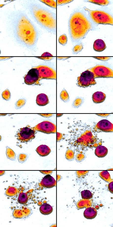
Apoptosis study: Acridine orange can be used to detect apoptotic cells, as cells undergoing apoptosis will show both DNA and RNA degradation.
Limitations of Acridine Orange Staining
While acridine orange staining is useful in many applications, it has some limitations:
Non-specific binding: Acridine orange can bind to other cellular components besides nucleic acids, which can sometimes result in background fluorescence. This can make it difficult to interpret the results accurately.
Limited to nucleic acids: This technique can only visualize DNA and RNA, so it doesn’t provide any information about other important cellular components, such as proteins or lipids.
Requires specialized equipment: A fluorescence microscope with UV light is essential to view the stained sample. This equipment can be expensive and may not be available in all laboratories.
Fading of fluorescence: The fluorescence produced by acridine orange can fade over time. This makes it difficult to store slides for long-term analysis, and the results must often be viewed immediately after staining.
Sample damage: UV light used in fluorescence microscopy can cause damage to the sample over time. Prolonged exposure can affect the accuracy of the results, particularly if trying to observe dynamic processes within living cells.
Distinguishing live and dead cells: Although acridine orange can differentiate between DNA and RNA, it doesn’t always distinguish live from dead cells effectively. Other complementary stains may be needed to confirm the viability of the cells.
Conclusion
Acridine orange staining is a powerful technique for studying DNA and RNA in cells. It is relatively easy to perform and provides valuable insights into cell health, apoptosis, and the presence of infections. By emitting different colors when binding to DNA or RNA, acridine orange helps researchers and doctors visualize what’s happening inside cells under a microscope.
However, like any technique, it has its limitations. Non-specific staining and the need for specialized equipment are notable challenges. Despite these drawbacks, acridine orange staining remains an important tool in cell biology and medical diagnostics, helping to distinguish between different types of nucleic acids and providing a deeper understanding of cellular processes.
Frequently Asked Questions (FAQ)
what do you mean by apoptosi?
Apoptosis is a form of programmed cell death that occurs in multicellular organisms. It is a natural process where cells undergo a controlled, orderly death as part of growth, development, and maintaining the health of an organism.
Write about DNA and RNA ?
DNA (Deoxyribonucleic Acid) and RNA (Ribonucleic Acid) are two fundamental types of nucleic acids that play key roles in storing and transmitting genetic information in living organisms.
DNA (Deoxyribonucleic Acid)
Related Articles


