
Fluorescent in-situ hybridization (FISH) is a versatile and effective method for identifying and localizing particular nucleic acid sequences in diploid cells. FISH is extremely useful in clinical diagnostics, genetic research, and cell biology because it provides precise spatial and temporal information on gene expression, chromosomal abnormalities, and genomic organization. To use FISH successfully, each step must be carefully optimized, from sample preparation and probe design to hybridization and imaging.
Table of Contents
Fluorescent In-Situ Hybridization
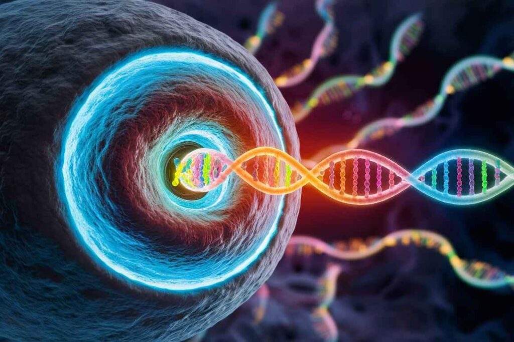
Fluorescent in-situ hybridization (FISH) is a strong molecular technique for detecting and localizing specific nucleic acid sequences within cells and tissues. This approach uses fluorescent probes that bind to complementary DNA or RNA sequences, allowing for viewing under a fluorescence microscope. FISH is used extensively in clinical diagnostics, genetic research, and cell biology to investigate chromosomal abnormalities, gene expression, and genome organization.
Principle of Fluorescent In-Situ Hybridization (FISH)
FISH involves the following key steps:
- Cells and tissues are fixed and permeabilized to give probe access to the target nucleic acids.
- Fluorescently labeled probes are hybridized with their complementary DNA or RNA sequences within the cell.
- Hybridized probes are identified and observed using fluorescence microscopy.
Process of Fluorescent In-Situ Hybridization (FISH) on Diploid Cells
1. Sample Preparation
Take diploid cells from the desired source (e.g., blood, cultured cells, tissue samples).
Prepare cell cultures on microscope slides or tissue slices.
2. Permeability and fixation
To maintain cellular architecture, fix the cells or tissue slices with methanol/acetic acid or formaldehyde.
Use a detergent like Triton X-100 to permeate the cells, allowing probe penetration.
3. Denaturation:
Denaturate the cells (with formamide or heat) to split the DNA strands and make the target sequences accessible to the probes.
4. Hybridization:
Denature the cells and then apply fluorescently labeled DNA or RNA probes.
Incubate at conditions that allow the probes to hybridize with their complementary target sequences (usually 37-42°C in a humid room).
5. Washes used after hybridization:
Wash the cells to eliminate loose and nonspecifically bound probes, often in a succession of more strict washes.
6. Counterstaining:
To see the cell nuclei and chromosome structure, stain the cells with a DNA-specific dye, such as DAPI (4′,6-diamidino-2-phenylindole).
7. Mounting:
To retain the fluorescence, put the slides in an anti-fade mounting medium.
8. Imaging:
Use a fluorescence microscope with the proper filters for the fluorophores to see the hybridized probes.
Take pictures and examine the location and intensity of the fluorescent signals.
Applications of Fluorescent In-Situ Hybridization (FISH)
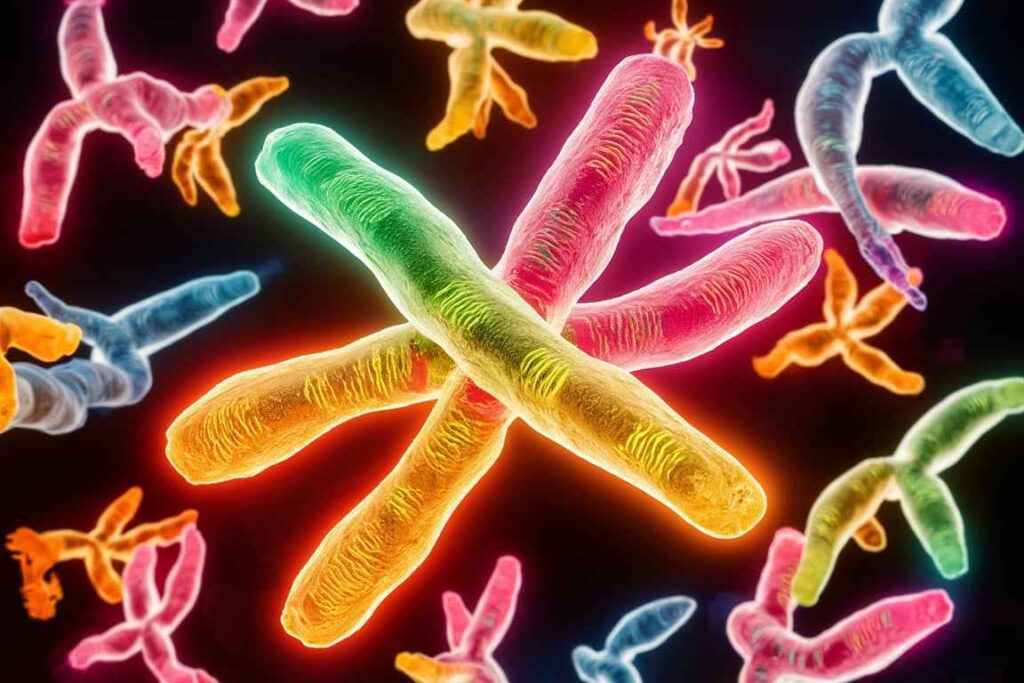
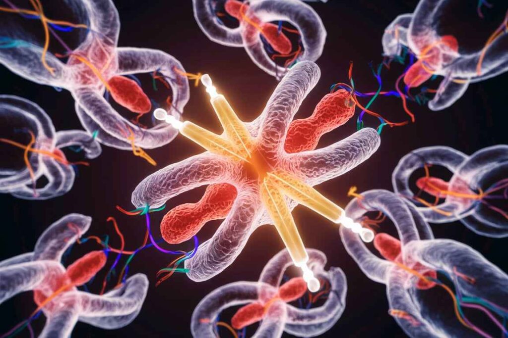
Chromosomal Aberrations:
Detecting structural chromosomal abnormalities such as translocations, deletions, duplications, and inversions.
Commonly used in cancer diagnostics to identify specific genetic alterations.
Gene Mapping:
Locating genes or genetic markers on chromosomes to study genetic linkage and genome organization.
Gene Expression:
Analyzing the expression patterns of specific genes by hybridizing probes to RNA transcripts in cells or tissues.
Prenatal Diagnosis:
Detecting chromosomal abnormalities in fetal cells obtained through amniocentesis or chorionic villus sampling.
Microbial Identification:
Identifying specific microbial species or strains in clinical samples by hybridizing probes to unique genetic sequences.
Example Protocol for Fluorescent In-Situ Hybridization (FISH) on Diploid Cells
Materials:
- Diploid cells (e.g., cultured fibroblasts)
- Fixative (e.g., methanol/acetic acid)
- Permeabilization buffer (e.g., PBS with 0.1% Triton X-100)
- Denaturation solution (e.g., 70% formamide in 2x SSC)
- Fluorescently labeled DNA probes
- Hybridization buffer (e.g., formamide, dextran sulfate, SSC)
- Wash buffers (e.g., 2x SSC, 0.1x SSC)
- Counterstain (e.g., DAPI)
- Anti-fade mounting medium
- Fluorescence microscope
Precautions and Considerations
1. Probe Design and Labeling:
Make that the probes are unique to the target sequences and tagged with the proper fluorophores.
Use controls to ensure probe specificity and hybridization efficiency.
2. Denaturation Conditions:
Optimize denaturation conditions to obtain single-stranded DNA without causing significant DNA breakdown.
3. Hybridisation Stringency:
Adjust the hybridization and wash conditions to strike a compromise between probe specificity and binding efficiency.
High stringency conditions (such as high temperature and low salt concentration) minimize non-specific binding.
4. Fluorescence Imaging:
To detect fluorophores, use a fluorescence microscope equipped with the appropriate filter sets and settings.
To avoid photobleaching, reduce light exposure and use anti-fade mounting media.
Frequently Asked Question
What is Fluorescent In-Situ Hybridization ?
Fluorescent in situ hybridization (FISH) is a strong molecular technique for detecting and localizing specific nucleic acid sequences within cells and tissues. This approach uses fluorescent probes that bind to complementary DNA or RNA sequences, allowing for viewing under a fluorescence microscope. FISH is used extensively in clinical diagnostics, genetic research, and cell biology to investigate chromosomal abnormalities, gene expression, and genome organization.
What are the applications of Fluorescent In-Situ Hybridization (FISH) ?
The applications of Fluorescent In-Situ Hybridization are
1. Chromosomal Aberrations
2. Gene Mapping
3. Gene Expression
4. Prenatal Diagnosis
5. Microbial Identification
Related Article