Microorganisms are all around us, but they are too small to be seen with the naked eye. Even under a microscope, many of them are colorless and transparent, which makes it hard to study their shape, size, and structure. This is where stains or dyes play a very important role in microbiology. They help color the microorganisms or specific parts of them, making them easier to see and understand.
What Are Stains or Dyes in Microbiology?
In microbiology, stains (also called dyes) are chemical substances used to give color to microorganisms or their internal structures. They don’t just make microbes look colorful they also help scientists study how cells are built, how they function, and how to identify different types of bacteria, fungi, or other microbes.
Summary of Stains or Dyes
- Stains or dyes are chemicals used in microbiology to color microorganisms, making them visible under a microscope.
- They help in identifying and studying microbes through different types like simple, differential (e.g., Gram stain), special, and negative stains.
- Staining works by chemical interaction between the dye and microbial cell surface, revealing key features for classification and diagnosis.
Table of Contents
Composition of Stains or Dyes
Most microbiological stains are made of three main components:
- Chromophore: This is the part of the molecule that gives the dye its color. It absorbs light and reflects a specific wavelength, which appears as a color to our eyes.
- Auxochrome: This is the part that binds the dye to the microorganism. It doesn’t give color, but it helps the stain stick to the cell by giving the molecule a positive or negative charge.
- Solvent: The dye is usually dissolved in water, alcohol, or another liquid, so it can be applied easily.
So, the stain has color (chromophore), stickiness (auxochrome), and a carrier (solvent) all working together to color the microbial cells.

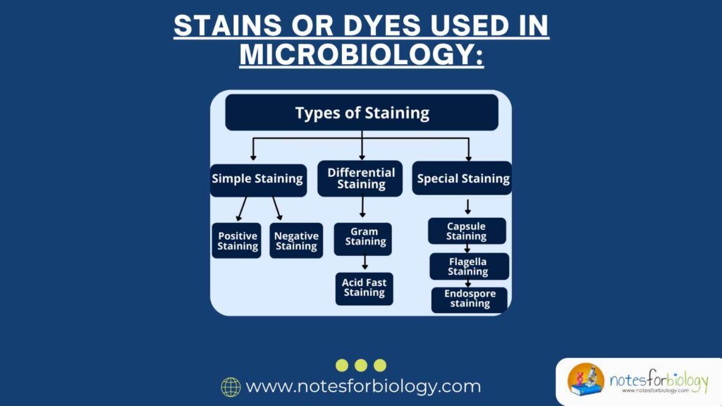
Types of Stains ( or Dyes) Used in Microbiology
There are several types of stains, and each has its own purpose. Let’s understand the major ones in a simple way:
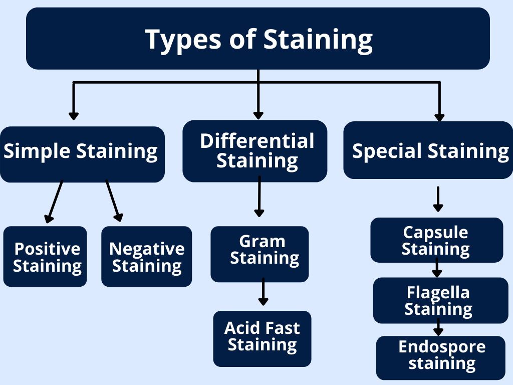
1. Simple Stains
- These stains use only one dyes.
- They are used to highlight the whole microorganism.
- They help us see basic features like shape, size, and arrangement.
- Examples: Methylene blue, Crystal violet, Safranin.
- How it works: The dyes bind to the cell and gives it a uniform color.
2. Differential Stains
- These stains use two or more dyes to differentiate between types of microorganisms.
- They help tell the difference between two groups of bacteria based on their cell wall structure.
The most common differential stains:
a. Gram Staining
- Divides bacteria into Gram-positive (purple) and Gram-negative (pink).
- Based on the thickness of the bacterial cell wall.
b. Acid-Fast Staining
- Used to identify bacteria like Mycobacterium (e.g., TB bacteria), which have waxy cell walls.
- Acid-fast bacteria remain red; others turn blue.
3. Special Stains
- These are used to color specific structures inside or outside the microorganism.
- They are like zooming in on a specific part of the cell.
Examples:
- Capsule stain – shows the protective capsule around bacteria.
- Endospore stain – colors tough spores formed by some bacteria.
- Flagella stain – highlights the tail-like structure used for movement.
4. Negative Stains
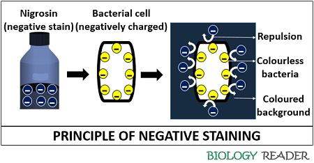
- These don’t stain the actual microorganism.
- Instead, they stain the background, making the microbe appear clear and bright.
Example: India ink or Nigrosin.
This is helpful for delicate structures like capsules, which might get damaged by harsh staining
Mechanism of Staining : How Do Stains Work?
Let’s understand how stains stick to microbes. It’s a little like magnets and glue!
Microbial cells carry either a positive or negative charge on their surface. Most bacterial cells are negatively charged. So, if the stain is positively charged (called a basic dye), it will be attracted to the negatively charged cell just like a magnet.
1. Basic Stain
- These have positive charges.
- They bind easily to negatively charged bacterial surfaces.
- Commonly used in microbiology.
Examples: Methylene blue, Crystal violet.
2. Acidic Stains
- These have negative charges.
- They are repelled by bacterial surfaces and stain the background instead.
Example: Eosin, Nigrosin.
3. Heat-Fixing
Before applying many stains, microbes are usually heat-fixed to a slide. This means they are gently passed through a flame. This step:
- Kills the microbe,
- Sticks it to the glass,
- And prepares it to absorb the stain better.
4. Staining Procedure
Each stain has its own steps. For example:
Gram Staining:
- Apply crystal violet (primary stain).
- Add iodine (fixes the dye).
- Wash with alcohol (decolorizes).
- Add safranin (counterstain).
Why Are Stains Important in Microbiology?
Stains are important in Microbiology,
- Improved Visibility: Makes tiny microbes visible under the microscope.
- Identification: Helps differentiate types of bacteria and other microbes.
- Structure Detection: Allows observation of capsules, spores, flagella, etc.
- Disease Diagnosis: Helps doctors detect bacteria in patient samples.
- Research and Learning: Crucial in education and scientific study.
Conclusion
Staining is one of the most important techniques in microbiology. It allows us to see and understand microorganisms that would otherwise be invisible. Whether it’s a simple dye that colors the whole cell, or a special stain that highlights tiny structures, these stains open a colorful window into the world of microbes.
By knowing the composition, types, and mechanism of staining, scientists and students can study bacteria and other microorganisms in great detail. These colorful tools make it possible to explore the hidden beauty and complexity of the microscopic world—one cell at a time.
Frequently Asked Questions (FAQs)
What are stains or dyes in microbiology?
Stains or dyes are chemical substances used to color microorganisms or their components, enhancing visibility under a microscope. They help differentiate between various microbial structures and types.
Why is staining important in microbiology?
Staining increases the contrast between microorganisms and their background, making them easier to observe and identify. It aids in studying cell morphology, structure, and classification.
How does Gram staining differentiate bacteria?
Gram staining differentiates bacteria based on their cell wall composition:
Gram-positive bacteria: Thick peptidoglycan layer; retain crystal violet stain and appear purple.
Gram-negative bacteria: Thin peptidoglycan layer; do not retain crystal violet but take up the counterstain (safranin) and appear pink.
Related Articles