Introduction
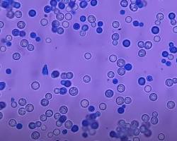
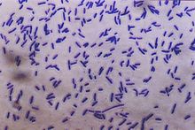
Simple staining is a fundamental method in microbiology that is used to color bacteria, making them more apparent when viewed under a microscope. In contrast to more intricate staining techniques (such as Gram staining), simple staining utilizes just a single dye to emphasize bacterial cells, facilitating the observation of their shape, size, and arrangement. It’s a quick and simple technique frequently employed in laboratories to examine microorganisms.
Table of Contents
Common Stains Used in Simple Staining
The common stains used in Simple Staining are:
Methylene Blue: A widely utilized dye. It’s affordable and simple to utilize.

Crystal Violet: Often utilized in basic staining, yet it is predominantly observed in Gram staining.

Safranin: Although it is usually employed as a counterstain in Gram staining, it may also be utilized by itself for straightforward staining.

Carbol Fuchsin: A vibrant pink/red dye that is used infrequently but remains an option for basic stains.
Materials Needed for Simple Staining
Microscope: For observing the colored bacteria.
Glass slides The surface for placing your bacterial sample.
Bunsen burner: To heat and adhere the bacteria to the slide.
Inoculation loop: For transferring the bacterial culture onto the slide.
Bacterial culture: A specimen of bacteria you wish to study (from an agar plate or liquid medium).
Stain: A specific dye used to tint the bacteria (for instance, methylene blue, crystal violet, or safranin).
Water: To rinse the slide.
Dropper: For dispensing water and staining onto the slide.
Paper towels or absorbent paper: To dry the slide following rinsing.
Step-by-Step Procedure for Simple Staining
Prepare the slide
- Use a clear glass slide.
- Put a tiny droplet of water (when using a solid culture) on the slide.
- Utilize an inoculation loop to place a small quantity of bacterial culture onto the slide.
- Distribute the culture uniformly to create a light layer.
- Let the smear dry in the air fully. Take your time with this step!
- Heat-fix the smear by passing the slide through a flame 2-3 times to eliminate the bacteria and ensure they adhere to the slide.
Prepare a Bacterial Smear
- For an agar plate: flame-sterilize your inoculating loop and allow it to cool. Pick a small amount of bacterial culture and spread it thinly over the water drop to make an even smear.
- For a liquid culture: Place a drop of the liquid culture directly onto the slide.
Let the smear dry
- Allow the smear to air dry for a couple of minutes. Exercise caution to avoid excess—allow it to dry naturally instead of applying excessive heat. If the smear is moist, it could come off when you attempt to stain it.
Heat Set the Smear (Optional but Suggested)
- After the smear is dry, you must “fix” the bacteria onto the slide. This is accomplished by methodically passing the slide 2-3 times through the flame of a Bunsen burner.
- The heat will eliminate the bacteria, making them adhere to the slide. Take care to avoid overheating the slide, since this may distort or harm the bacterial cells.
Using the stain
- With your slide now prepared, proceed to apply the stain (such as methylene blue, crystal violet, or safranin). Only a couple of drops are needed.
- Allow the stain to rest on the slide for approximately 30 seconds to 1 minute. Be patient; this is when the bacteria take in the dye.
Rinse the Slide
- Following the staining period, carefully rinse the slide with distilled water to remove any surplus dye. Tilt the slide to prevent disrupting the smear.
Blot Dry
- Gently dab the slide using paper towels or filter paper to eliminate any surplus water. Refrain from rubbing the slide, since this may disturb the smear.
Examine Under the Microscope
- Position the slide on the stage of the microscope. Begin with the lowest power (typically 10x) to find the bacterial cells.
- After locating the smear, change to a higher magnification (100x with oil immersion) to observe the details distinctly.
- The bacteria need to be dyed and seen as colored cells on a light background.
Importance of Simple Staining Technique
- Examination of Form and Organization: It aids in recognizing the shape of bacteria (such as cocci, bacilli, or spirals) and their arrangement (e.g., in chains, clusters, or pairs).
- Fast and Simple: This method needs minimal prep and doesn’t involve various stains, making it perfect for novices or for a rapid inspection of a bacterial culture.
- Preparation for advanced techniques: Having a grasp of basic simple staining aids in comprehending more intricate staining procedures, such as Gram staining, which offers more in-depth insights into bacterial traits.
Advantages of Simple Staining
- Easy to Execute: Basic staining needs minimal chemicals and time. You just require a single stain, simplifying the learning process for newcomers.
- Fundamental Cell Morphology: It enables the observation of fundamental cell forms (cocci, bacilli, spirilla) and arrangements (clusters, chains).
- Fast Outcomes: The whole procedure can be completed in under 30 minutes, which is advantageous when you need to swiftly analyze bacteria.
- Economical: The procedure needs few reagents and tools, rendering it a cost-saving approach.
- Flexibility: It is applicable for staining different types of microorganisms, such as bacteria, fungi, and certain protozoa.
Drawbacks of Basic Staining
- Restricted Information: Basic staining offers minimal insights into other crucial bacterial traits (e.g., movement, spore presence, or cell wall structure).
- Challenges in Recognizing Similar Organisms: It can be difficult to tell apart closely related bacterial species that possess similar morphological traits.
- Risk of Overstaining: Prolonged staining duration can result in overstaining, hindering the visibility of cellular details and complicating the accurate observation of bacterial structure.
- Risk of Overheating: Excessive heat during slide fixation may warp the bacterial cells, affecting the quality of microscopic examination.
Frequent Errors in Basic Staining
- Not Allowing the Smear to Dry: If the smear remains wet, it may wash away when you apply the stain. Always allow it to dry fully before applying stain.
- Excessive Heating of the Slide: Overheating the slide excessively may harm the bacteria. Simply move the slide through the flame swiftly, avoiding excess.
- Applying Excessive Stain: Just a couple of drops of dye are sufficient. Excessive use can hinder visibility of the bacteria or lead to the smear washing away.
- Not Rinsing Adequately: Wash away the excess stain. Excess dye remaining on the slide can obscure the visibility of the bacteria, while insufficient rinsing may result in a smear that is too dark for clear observation.
Conclusion
Simple staining is an efficient and straightforward technique to observe bacteria using a microscope. It aids in determining their form, dimensions, and organization, making it a useful initial step in examining microorganisms. Although it lacks the detailed insights of Gram staining, it serves as a valuable method for novices and an excellent foundation in microbiology. By using a single stain, you can obtain important information about the fundamental traits of bacterial cells.
Frequently Asked Question (FAQ)
What is the difference between simple staining and Gram staining?
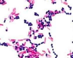
Simple staining employs a single dye to color bacteria, while Gram staining utilizes two distinct dyes to categorize bacteria as Gram-positive or Gram-negative depending on the composition of their cell walls. Basic shapes and arrangements are revealed through simple staining, whereas Gram staining offers more comprehensive details regarding bacterial types.
Why is heat fixing important in simple staining?
Heat fixing is essential as it eliminates bacteria, maintains their structure, and assists in adhering them to the slide. If heat fixing is not applied, the bacteria could rinse away during staining or fail to absorb the stain adequately.
What happens if I use too much stain?
Applying excessive stain can obscure the visibility of the bacteria. The smear could turn overly dark, making it difficult to distinguish the bacteria from the background. Always apply a few drops of stain for optimal contrast.
Related Articles


