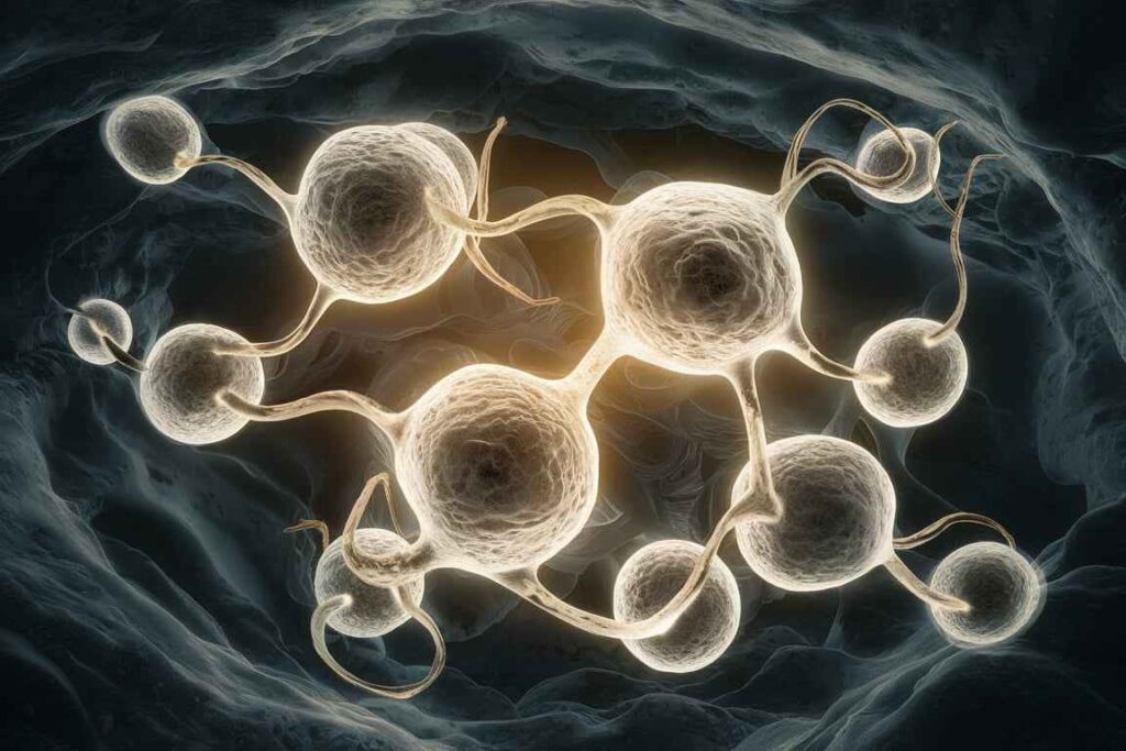
Primary cell culture refers to the process of isolating and culturing cells directly from a living organism or tissue sample. Primary cultures are valuable tools for studying cell behavior, function, and interactions in a controlled laboratory environment.
Table of Contents
Primary cell culture
Primary cell culture refers to the process of isolating cells from a tissue source (such as an animal or plant) and maintaining them in an artificial environment to allow for growth and study. These cells are directly obtained from living tissue and reflect the physiological state of the tissue more accurately than established cell lines.
Materials & Reagents
- Fertile chicken eggs (10-11 day-old embryos)
- Ethanol (70%
- Phosphate buffered saline (PBS), sterile
- Trypsin-EDTA solution (0.25% trypsin, 0.02% EDTA)
- Dulbecco’s Modified Eagle Medium (DMEM) or the Minimum Essential Medium (MEM)
- Fetal Bovine Serum (FBS)
- Antibiotics (Penicillin-Streptomycin
- Use sterilized Petri dishes and Hank’s Balanced Salt Solution (HBSS).
- Conical tubes (15 and 50 mL)
- Cell Culture Flasks (T-25 or T-75)
- Scalpels, forceps, and scissors.
- Pipettes and Pipette Tips
- Laminar flow hood with CO2 incubator (37°C, 5% CO2).
- Centrifuge
Protocol:
Preparation and Sterilization
Ensure that all chemicals, equipment, and surfaces are sterile.
Clean the laminar flow hood with 70% ethanol.
Pre-heat DMEM/MEM and other solutions to 37°C.
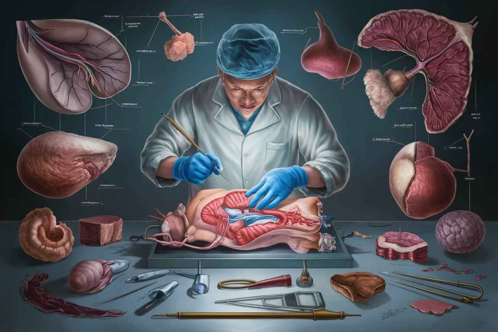
Harvesting chick embryos
Disinfect the eggshell’s surface with 70% ethanol.
Crack the egg and carefully transfer the embryo to a sterile Petri plate with PBS.
Dissection and Tissue Collection.
Using sterile forceps and scissors, detach the embryo’s head and internal organs while keeping the body intact.
Cut the embryo body into little pieces (~1 mm) with sterile scalpels.
Enzymatic digestion
Transfer the minced tissue to a conical tube with 5-10 mL of trypsin-EDTA solution.
Incubate at 37°C for 10-15 minutes, gently swirling to assist digestion.
Following digestion, add an equal amount of complete medium (DMEM/MEM supplemented with 10% FBS and antibiotics) to neutralize trypsin.
Mechanical Dissociation
Pipette gently up and down to further separate the cells and create a single-cell suspension.
To eliminate undigested tissue, pass the suspension through a sterile cell strainer or gauze.
Centrifugation
To pellet the cells, centrifuge the suspension at 1,000 rpm at room temperature for 5 minutes.
Remove the supernatant and re-suspend the cell pellet in fresh complete media.
Seeding Cells
Count cells with a hemocytometer and adjust density to 1-2 × 10^5 cells/ml.
Seed the cells into culture flasks containing the required volume of complete media.
Place the flasks in a CO2 incubator set to 37°C with 5% CO2.
Culture Maintenance
Every 2-3 days, change the medium by carefully removing the old medium and replacing it with fresh, pre-warmed complete media.
Examine cell growth and morphology with a microscope. CEFs normally adhere within 24 hours and begin to proliferate.
Subculturing
Once cells achieve 70-80% confluence, they can be subcultured:
Remove the media and wash the cells with PBS.
To detach the cells, add a trypsin-EDTA solution and incubate for 2–5 minutes at 37°C.
Neutralize trypsin in complete medium, then collect the cells and centrifuge at 1,000 rpm for 5 minutes.
Resuspend the cell pellet in fresh complete media and seed it in new culture flasks.
Notes
Sterility: Maintain sterility throughout the procedure to avoid contamination.
Incubation Conditions: Maintain a consistent temperature and CO2 levels in the incubator.
Cell viability: Use trypan blue exclusion method to assess cell viability before seeding.
Ethical Considerations: Follow ethical guidelines and regulations for the use of animal embryos in research.
Frequently Asked Question
Define Primary cell culture.
Primary cell culture refers to the process of isolating cells from a tissue source (such as an animal or plant) and maintaining them in an artificial environment to allow for growth and study. These cells are directly obtained from living tissue and reflect the physiological state of the tissue more accurately than established cell lines.
What are the key considerations of Primary cell culture ?
The key considerations of Primary cell culture are
1. Sterility:
2. Cell Source
3. Medium:
4. Environment
Related Article
Salmonella: morphology, antigenic structure, cultural and biochemical characteristics