Different degrees of research and study of the body’s muscles are referred to by the names Muscle-skeletal Muscle-gross and Ultrastructure. An explanation of each is provided below:
Table of Contents
Muscle-Skeletal System

The human body’s structure, support, and mobility are provided by the intricate network of bones, joints, muscles, tendons, and ligaments that make up the musculoskeletal system.
It makes it possible for us to carry out everyday tasks like lifting and carrying things as well as jogging and walking.
Components of the Muscle-Skeletal System
1. Bones
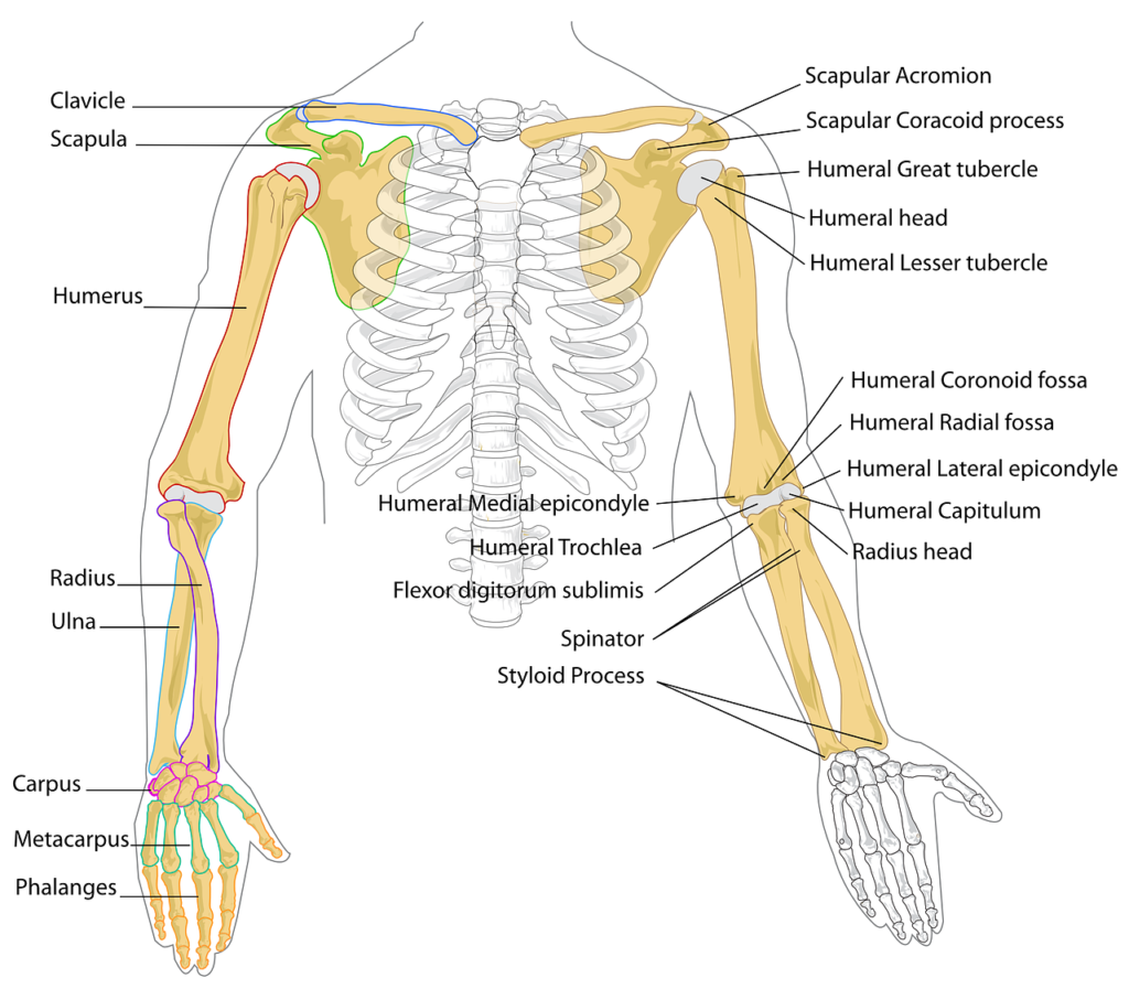
The body is supported, protected, and has leverage for movement thanks to the bones that make up the muscle-skeletal system. The adult human body contains 206 bones, which are arranged according to their structure and use. Here is a summary:
Types of Bones:
- Long bones are made for support and mobility; they are longer than they are wide.
- The femur, tibia, humerus, radius, and ulna are examples of long bones in the human body.
- The short bones, which are roughly cube-shaped, offer support and stability.
- Examples of short bones in the human body are the tarsal and carpal bones.
- Thin, flat, and frequently curved, flat bones offer surface area for muscle attachment and protect internal organs.
- Examples of flat bones in the human body are the scapula, ribs, sternum, and skull.
- Irregular bones are intricate structures with distinct roles.
- Vertebrae, face bones, and certain pelvic bones are examples of irregular bones in the human body.
- Sesamoid bones are tiny, flat bones that are inserted into tendons to protect them and change the angle at which they pull.
- Examples of sesamoid bones in the human body are the patella (kneecap) and the hands and feet.
Structure and Function
- Provide the body’s inflexible skeleton.
- Safeguard essential organs (e.g., ribs protect the heart and lungs, skull protects the brain).
- Keep vital minerals like phosphorus and calcium in storage.
- include bone marrow, which is used to make blood cells.
2. Muscles
The contractile tissues that provide force, allowing for posture maintenance and movement are muscles.The musculoskeletal system relies heavily on muscles to provide movement, stability, and support.
There are over 600 muscles in the human body, categorized into three main types:
Types
- Skeletal muscles are striated, voluntary muscles that are connected to bones and are in charge of posture and movement.
- Involuntary smooth muscles are present in the walls of organs, such as the stomach and blood arteries.
- The heart’s striated, involuntary muscle is called the cardiac muscle.
Human Muscle Anatomy
- Tendons: Sturdy connective tissues that join bones and muscles.
- Ligaments are pliable connective tissues that stabilize joints by joining bones.
Functions:
- Movement: We can walk, run, jump, and engage in other activities because our muscles contract and relax to create movement.
- Posture: By bearing the weight of the body and keeping us upright, muscles aid in maintaining good posture.
- Stability: Joints are shielded from harm by muscles that stabilize them and limit excessive movement.
- Heat Generation: The contraction of muscles produces heat, which aids in regulating body temperature.
- Protection: By supporting and cushioning interior organs, muscles offer protection.
3. Joints
Joints are the places where two or more bones meet, allowing for movement and flexibility in the musculoskeletal system.
They are essential for various activities, from simple gestures to complex athletic movements.
Types:
- Fibrous joints are immobile because they firmly keep bones together, enabling little to no mobility. Examples include the joints (sutures) that connect the skull’s bones.
- Cartilaginous joints are somewhat movable and are frequently seen in situations requiring stability and flexibility. Examples are the joints between the ribs and the sternum and the intervertebral discs, which connect the vertebrae.
- Synovial joints are freely movable and have a large range of motion. They are distinguished by the presence of cartilage, synovial fluid, and a joint capsule. Example: Knee, Shoulder
Functions :
- Movement: From basic gestures to intricate sports movements, joints allow for a vast range of motion. They serve as pivots, enabling muscles to generate motion and apply force.
- Stability: By keeping bones together, joints give the skeletal system stability. Maintaining posture and safeguarding internal organs depend on this stability.
- Shock Absorption: During exercises like running and jumping, some joints, like the knee and ankle, reduce the force applied to the body by acting as shock absorbers.
- Weight Bearing: The ability to stand, walk, and run is made possible by the lower limb joints, such as the hip and knee, which support the body’s weight.
- Growth: In children, the head can grow and expand thanks to specific joints, especially those that connect the skull’s bones.
4. Cartilage
One specialized connective tissue that is essential to the musculoskeletal system is cartilage.
It is a durable, supple, and smooth tissue that gives different bodily components flexibility, support, and defense.
Key Functions of Cartilage:
- Cartilage reduces the impact of forces on joints during movement by acting as a shock absorber. In weight-bearing joints like the knees and hips, this is especially crucial.
- Smooth Surface for Movement: Because cartilage has a smooth surface, there is less friction between bones, which makes movement painless and easy.
- Support and Structure: The ears, nose, and trachea are just a few of the body parts that cartilage supports structurally. It also aids in keeping some organs in their proper form.
- Flexibility: A large range of motion is made possible by cartilage’s ability to give joints flexibility. This is particularly crucial for joints like the spine and shoulder.
Types of Cartilage:
- Hyaline Cartilage: The most prevalent kind, which is present in the nose, trachea, ribs, and at the extremities of bones in joints. For structural support and joint mobility, it offers smooth surfaces.
- Strong and resilient, fibrocartilage is present in the pubic symphysis, the menisci of the knee joint, and intervertebral discs. It offers both stress absorption and support.
- Found in the outer ear, epiglottis, and portions of the larynx, elastic cartilage is pliable and elastic.
- It offers assistance and flexibility.
Functions of Muscle-Skeletal System
- Support and Structure: Gives the body the structure it needs to keep its posture and form.
- Movement: Facilitates a variety of motions, including large motor movements and fine motor skills.
- Protection: Prevents harm to critical organs such as the brain, heart, and lungs.
- Production of Blood Cells: The bone marrow found in bones is responsible for the production of blood cells.
- Mineral Storage: Holds vital minerals such as phosphorus and calcium.
- Heat Generation: The contraction of muscles produces heat, which aids in regulating body temperature.
In conclusion, because it enables us to move, operate, and thrive, the musculoskeletal system is critical to our general health and well-being.
Muscle-Gross Structure
The study of bodily structures that are apparent to the unaided eye is known as gross anatomy.
Examining the general size, shape, and distribution of muscles inside the body is known as gross structure.
Functions of Muscle-Skeletal System
The main components of muscle gross structure are as follows:
Muscle Fiber Types:
Different kinds of muscle fibers, each with unique properties and purposes, make up muscles:
1. Type I (Slow-Twitch) Fibers:
- Specifically designed for endurance exercises
- Rich in mitochondria and myoglobin
- ATP is produced via aerobic metabolism.
- Reduced contraction rate
- Able to withstand fatigue
2. Type IIa (Fast-Twitch Oxidative) Fibers:
- Intermediate fibers that combine Type I and Type IIb properties
- Moderate fatigue resistance
- moderate pace of contraction
- Both aerobic and anaerobic metabolism are possible.
3.Type IIb (Fast-Twitch Glycolytic) Fibers:
- Specialized in speed and power
- Low levels of mitochondria and myoglobin
- Utilize anaerobic glycolysis to produce ATP.
- Increased contraction speed
- Fatigue rapidly
Muscle Architecture
The arrangement of muscle fibers within a muscle can influence its function:
1. Parallel Muscles:
One kind of skeletal muscle is a parallel muscle, which has fibers that run parallel to its length, frequently between its insertion and origin places.
- Parallel to the muscle’s long axis are fibers.
- Produce force across a considerable distance.
- Sartorius and rectus abdominis are two examples.
2. Fusiform Muscles:
Fusiform muscles are a kind of skeletal muscle that resembles a spindle and is distinguished by its tapered ends and protruding center, or belly.
- Fibers that converge at both ends form a spindle.
- Range of motion and force balance
- Examples are the brachialis and brachii biceps.
3. Pennate Muscles:
The skeletal muscle type known as pennate muscles is distinguished by its feather-like fiber arrangement, in which fibers obliquely connect to a central tendon.
- The arrangement of the fibers is perpendicular to the tendon.
- Produce more force because there are more fibers.
- Unipennate, bipennate, and multipennate types
- For instance, the deltoid and rectus femoris
Muscle Attachments
- Origin: A muscle’s fixed place of attachment.
- Insertion: A muscle’s moveable point of attachment.
- Action: The particular motion brought about by a contraction of the muscles.
Ultrastructure of Muscle
The microscopic and sub-microscopic arrangement of muscle tissue, especially at the cellular level, is referred to as the ultrastructure of muscle. Muscle fibers, the specialized cells that make up muscles, have particular characteristics that enable force production and contraction. Skeletal muscles are the finest place to study the ultrastructure because, when viewed under a microscope, they appear extremely ordered.
Key Components of Muscle Ultrastructure:
- Muscle fibers are composed of long, cylindrical structures called myofibrils.
- Sarcomeres: Made up of thick (myosin) and thin (actin) filaments, they are the fundamental contractile components of muscle.
- Actin and Myosin Filaments: These proteins work together via a sliding filament mechanism to produce muscle contraction.
- A system of tubules called the sarcoplasmic reticulum stores and releases calcium ions, which are necessary for muscle contraction.
Key Functions of Muscle Ultrastructure:
- Effective Contraction: Muscle contraction is made possible by the highly ordered structure of muscle fibers, which includes the arrangement of myofibrils, sarcomeres, and contractile proteins.
- Accurate Control: Fine motor abilities and coordinated motions are made possible by the exact control of muscle contraction provided by the complex network of T-tubules and the SR.
- Rapid Response: Muscle contraction and relaxation are made possible by the quick release of calcium ions from the SR and the quick conduction of nerve impulses through T-tubules.
- Energy Efficiency: The overall energy efficiency of muscle contraction is influenced by the configuration of myofibrils and the use of metabolic pathways that use less energy.
Conclusion
There are close connections between the muscle-skeletal system, muscle-gross structure, and muscle ultrastructure. A muscle’s ultrastructure allows it to contract and produce force, whereas its gross structure dictates how it functions. We can gain a greater understanding of the musculoskeletal system’s functioning and how many circumstances, like aging, illness, and injury, can impact it by investigating these levels of organization.
Frequently Asked Questions (FAQ)
What do you mean by Muscle-Skeletal System?
The human body’s structure, support, and mobility are provided by the intricate network of bones, joints, muscles, tendons, and ligaments that make up the musculoskeletal system.
Define Muscle-Gross Structure.
The study of bodily structures that are apparent to the unaided eye is known as gross anatomy.
Examining the general size, shape, and distribution of muscles inside the body is known as gross structure
What is the relationship between muscle-skeletal system, muscle-gross structure, and muscle ultrastructure?
There are close connections between the muscle-skeletal system, muscle-gross structure, and muscle ultrastructure. A muscle’s ultrastructure allows it to contract and produce force, whereas its gross structure dictates how it functions. We can gain a greater understanding of the musculoskeletal system’s functioning and how many circumstances, like aging, illness, and injury, can impact it by investigating these levels of organization.


