Kidneys are the essential organ in the human excretory system, primarily responsible for filtering blood, removing waste products, regulating electrolyte balance, and maintaining fluid homeostasis. Situated within the abdominal cavity, the kidneys perform multiple critical physiological functions necessary for life. Apart from excretion, they also contribute to blood pressure regulation, acid-base balance, and the production of hormones vital for red blood cell formation and bone health.
Table of Contents
Definition of Kidney
A kidney is a paired, bean-shaped organ present in vertebrates, including humans, which plays a central role in the regulation of various bodily processes. Its primary function is to remove nitrogenous waste from the blood and maintain the body’s internal environment through selective filtration and excretion. Besides excretory duties, kidneys also perform vital endocrine and regulatory functions, making them indispensable for survival.
Position and Size of Kidneys
The kidneys are located in the posterior part of the abdominal cavity, known as the retroperitoneal space. They are positioned on either side of the vertebral column, typically extending from the level of the twelfth thoracic vertebra (T12) to the third lumbar vertebra (L3). Interestingly, the right kidney lies slightly lower than the left due to the presence of the liver on its superior side.
Regarding their dimensions, each kidney measures approximately 10 to 12 centimeters in length, 5 to 7 centimeters in width, and about 2.5 to 3 centimeters in thickness. The weight of a single kidney in an average adult ranges between 120 and 170 grams, with slight variations based on age, gender, and body size.
External Structure of Kidneys
Renal Capsule
The outermost covering of the kidney is known as the renal capsule. It is a tough, fibrous, and transparent connective tissue layer that adheres firmly to the kidney’s surface. This capsule provides protection against physical injury and infections.
Adipose Capsule (Perirenal Fat)
Surrounding the renal capsule is a thick layer of adipose tissue called the adipose capsule or perirenal fat. This fatty layer acts as a cushioning pad, absorbing shocks and protecting the kidney from external trauma. Additionally, it plays a role in insulating the kidney and maintaining its position within the abdominal cavity.
Renal Fascia
External to the adipose capsule lies the renal fascia, a thin yet strong layer of connective tissue that encloses both the kidney and the surrounding fat. The renal fascia anchors the kidneys to nearby structures, preventing displacement during bodily movements.
Pararenal Fat
Outside the renal fascia is another layer of fat known as the pararenal fat. It further cushions the kidneys and acts as a secondary protective barrier, insulating the organ from mechanical injuries and temperature fluctuations.
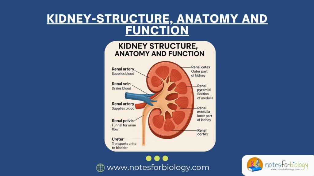
Renal Hilum
The medial border of each kidney contains a vertical slit-like opening known as the renal hilum. Through this hilum, several structures such as the renal artery, renal vein, lymphatic vessels, nerves, and ureter enter and exit the kidney. The hilum leads into a spacious cavity called the renal sinus, which houses the renal pelvis, calyces, blood vessels, and nerves.
Internal Structure of Kidneys
Renal Cortex
The outer region of the kidney is termed the renal cortex. It appears reddish-brown in color and possesses a granular texture due to the presence of numerous renal corpuscles. The renal cortex is primarily responsible for the initial phases of blood filtration as it houses the glomeruli and convoluted tubules.
Renal Medulla
Beneath the renal cortex lies the renal medulla. This inner region is darker and organized into several cone-shaped structures known as renal pyramids. Each pyramid consists of parallel bundles of urine-collecting tubules and blood vessels. The apex of each pyramid is called the renal papilla, which projects into a minor calyx.
Renal Columns
Intervening between the renal pyramids are extensions of cortical tissue known as renal columns. These columns contain blood vessels and connective tissue, and they provide structural support to the kidney while separating the adjacent pyramids.
Renal Pelvis
The innermost region of the kidney consists of the renal pelvis, a funnel-shaped cavity responsible for collecting urine produced by the nephrons. The pelvis is formed by the convergence of several minor calyces, which in turn merge into major calyces. From the renal pelvis, urine passes into the ureter for transportation to the urinary bladder.
Microscopic Structure of Kidneys
Nephrons
The fundamental structural and functional unit of the kidney is the nephron. Each kidney contains approximately one to one and a half million nephrons. These microscopic units are responsible for filtering blood, reabsorbing essential substances, and excreting waste products as urine.
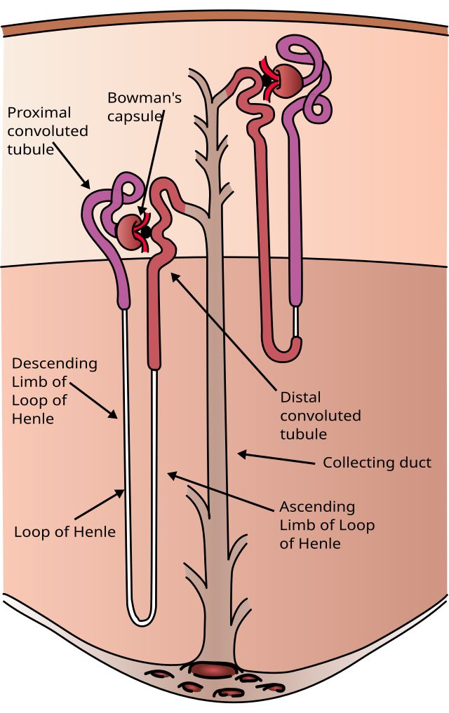
Renal Corpuscle
A nephron begins with the renal corpuscle, which is located in the renal cortex. The renal corpuscle consists of two main components: the glomerulus and the Bowman’s capsule. The glomerulus is a network of fenestrated capillaries through which blood is filtered. Surrounding the glomerulus is Bowman’s capsule, a double-walled, cup-shaped structure that collects the filtrate from the blood.
Renal Tubule
Extending from Bowman’s capsule is the renal tubule, a long, coiled tubular structure that processes the filtrate through reabsorption and secretion. The renal tubule comprises several segments: the proximal convoluted tubule (PCT), the loop of Henle, the distal convoluted tubule (DCT), and the collecting duct.
Juxtaglomerular Apparatus
Situated near the renal corpuscle is a specialized structure called the juxtaglomerular apparatus (JGA). This apparatus regulates the rate of glomerular filtration and helps control blood pressure by releasing the enzyme renin. It consists of juxtaglomerular cells, macula densa cells, and mesangial cells.
Blood Supply of Kidney
The kidneys receive a significant portion of the cardiac output, accounting for approximately 20 to 25 percent of the total blood pumped by the heart. Each kidney is supplied by a renal artery, which arises directly from the abdominal aorta.
Upon entering the kidney at the hilum, the renal artery branches into segmental arteries. These further divide into interlobar arteries that travel through the renal columns and reach the boundary between the cortex and medulla. Here, they form arcuate arteries, which curve along the base of the renal pyramids. From the arcuate arteries, smaller interlobular arteries arise and extend into the cortex, giving rise to afferent arterioles. These arterioles supply the glomerular capillaries, where filtration occurs. The blood exits the glomerulus through efferent arterioles, which then form peritubular capillaries and vasa recta, supplying oxygen and nutrients to the renal tissues.
Venous blood is drained by a network of interlobular, arcuate, interlobar, and segmental veins, eventually merging into the renal vein, which empties into the inferior vena cava.
Functions of Kidney
Filtration of Blood
One of the most important functions of the kidney is to filter blood, removing waste products such as urea, creatinine, and uric acid, along with excess ions and water. This filtration takes place in the glomerular capillaries and results in the formation of a protein-free filtrate.
Reabsorption
After filtration, the nephron selectively reabsorbs essential substances such as glucose, amino acids, sodium, potassium, calcium, and water from the tubular fluid back into the bloodstream. Most of this reabsorption occurs in the proximal convoluted tubule.
Secretion
It also actively secretes certain waste products, drugs, and excess ions into the renal tubules from the surrounding peritubular capillaries. This process ensures the elimination of substances that were not initially filtered at the glomerulus.
Excretion
The final urine, formed after reabsorption and secretion, contains waste materials and excess water. It is collected in the renal pelvis and transported to the urinary bladder via the ureter, from where it is eventually expelled from the body.
Regulation of Blood Pressure
It plays a key role in regulating blood pressure through the renin-angiotensin-aldosterone system (RAAS). In response to reduced blood flow or sodium levels, the juxtaglomerular cells secrete renin, which initiates a hormonal cascade to restore blood pressure and blood volume.
Regulation of Acid-Base Balance
By controlling the excretion of hydrogen ions and the reabsorption of bicarbonate, it maintains the acid-base balance of the blood. This regulation is crucial for maintaining the optimal pH required for enzymatic and metabolic activities.
Regulation of Electrolyte Balance
It regulates the concentration of various electrolytes such as sodium, potassium, calcium, and phosphate. This regulation is necessary for maintaining nerve impulse conduction, muscle contraction, and other physiological functions.
Erythropoiesis Regulation
In response to decreased oxygen levels in the blood, the kidney secretes the hormone erythropoietin. This hormone stimulates the bone marrow to produce red blood cells, ensuring adequate oxygen transport.
Activation of Vitamin D
It converts inactive vitamin D into its active form, calcitriol, which is essential for the absorption of calcium and phosphate from the intestines. This process is vital for maintaining bone health and mineral balance.
Conclusion
The kidney is a highly specialized and indispensable organ that performs a variety of excretory, regulatory, and endocrine functions. From filtering blood and removing waste to regulating electrolyte balance, blood pressure, and red blood cell production, the kidney’s functions are vital for sustaining life. Its complex structure, comprising external protective layers, internal regions, microscopic nephrons, and an elaborate blood supply, enables it to carry out these crucial roles efficiently. A comprehensive understanding of its anatomy and physiology is essential for appreciating its importance in maintaining the body’s internal environment.
Frequently Asked Questions (FAQ)
What is the main function of the kidney?
The main function of the kidney is to filter blood, remove waste products, and regulate fluid and electrolyte balance in the body.
Where are the kidneys located?
The kidneys are located in the upper abdominal cavity on either side of the vertebral column, between the T12 and L3 vertebrae.
What is a nephron?
A nephron is the microscopic functional unit of the kidney responsible for filtering blood and forming urine