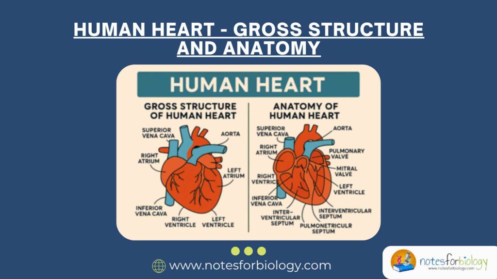
The human heart is a vital muscular organ that plays a central role in maintaining the circulation of blood throughout the body. It acts as a pump that keeps oxygenated blood flowing to various body tissues while collecting and sending deoxygenated blood to the lungs for purification. The heart can be described in terms of its gross structure and anatomy. The gross structure of the heart refers to its overall shape, size, position, coverings, and external features. The anatomy of the heart describes the detailed internal organization, including its chambers, valves, blood supply, and conducting system.
Summary of Human Heart
- The human heart is a muscular organ that pumps blood throughout the body.
- It has four chambers: two atria and two ventricles.
- Valves, coronary vessels, and a conducting system control blood flow and heartbeat.
Table of Contents
Gross Structure of the Human Heart
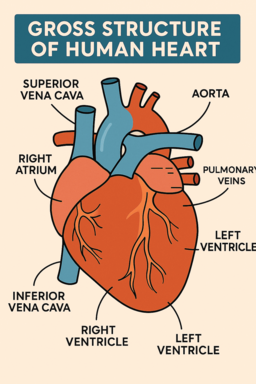
The heart is a hollow, muscular organ located in the thoracic cavity, slightly to the left of the midline. It is roughly the size of a fist and shaped like a cone. The heart has four chambers: two atria on top and two ventricles at the bottom. It is enclosed in a double-walled sac called the pericardium, which protects and anchors it. The heart pumps blood throughout the body, supplying oxygen and nutrients to tissues.
Shape, Size, and Position
The human heart has a cone-shaped structure and is a muscular, hollow organ. It typically weighs between 250 and 350 grams in adults. The heart is about the size of a closed human fist. It is positioned within the thoracic cavity, specifically in a region called the mediastinum, which lies between the two lungs.
It is located in such a way that its base, which is the broad upper part, is directed upward, backward, and to the right. The apex, which is the narrow, pointed lower end of the heart, is directed downward, forward, and to the left. The apex rests on the diaphragm at approximately the fifth intercostal space. Due to this arrangement, about two-thirds of the heart lies to the left of the midline of the body, while the remaining one-third lies to the right.
Coverings of the Heart (Pericardium)
The heart is covered by a protective membrane called the pericardium. This is a double-walled sac that serves to protect the human heart, hold it in place within the thoracic cavity, and prevent excessive movement. It also acts as a barrier to infection and reduces friction during the heart’s movements.
The pericardium consists of two main parts. The outer portion is known as the fibrous pericardium, which is a tough, dense, inelastic connective tissue layer. It is attached to the diaphragm, the posterior surface of the sternum, and the great vessels of the heart, thereby anchoring the heart securely within the chest.
The inner portion is called the serous pericardium, which is a thin, delicate membrane. It is further divided into two layers: the parietal layer, which lines the inner surface of the fibrous pericardium, and the visceral layer, also called the epicardium, which closely adheres to the external surface of the heart. Between these two layers lies a narrow space known as the pericardial cavity, which contains a small amount of pericardial fluid. This fluid functions as a lubricant, reducing friction as the heart beats.
External Features of the Human Heart
The external appearance of the heart reveals several important features that help in identifying its structure and function. The heart is divided externally into four chambers: two superior chambers known as the atria and two inferior chambers called the ventricles.
Each atrium possesses a small, ear-shaped appendage called an auricle. The auricles increase the capacity of the atria to accommodate more blood. Certain grooves or depressions are also visible on the outer surface of the human heart. These are known as sulci (singular: sulcus) and mark the boundaries between the chambers of the heart. The most prominent sulci include:
- The coronary sulcus, which encircles the heart horizontally and separates the atria from the ventricles.
- The anterior interventricular sulcus, which is found on the anterior surface of the heart and marks the boundary between the right and left ventricles.
- The posterior interventricular sulcus, which is present on the posterior surface and similarly divides the two ventricles.
These sulci also serve as pathways for the major blood vessels of the human heart, including the coronary arteries and cardiac veins.
Anatomy of the Human Heart (Internal Structure)
Chambers of the Heart
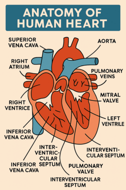
The human heart is internally divided into four chambers. These are the right atrium, right ventricle, left atrium, and left ventricle. The right atrium receives deoxygenated blood from the body through the superior vena cava, inferior vena cava, and coronary sinus. Its inner surface is marked by muscular ridges known as pectinate muscles. The right ventricle receives this deoxygenated blood from the right atrium and pumps it into the lungs through the pulmonary trunk, which divides into the right and left pulmonary arteries. Its inner surface is lined with irregular muscular ridges called trabeculae carneae and contains papillary muscles attached to fibrous cords known as chordae tendineae, which help control the heart valves.
The left atrium receives oxygenated blood from the lungs through four pulmonary veins. Its inner surface is smooth, except in the region of the auricle where pectinate muscles are found. The left ventricle, which has the thickest muscular wall among the four chambers, receives oxygenated blood from the left atrium and pumps it into the aorta for distribution to the entire body. Like the right ventricle, it also contains trabeculae carneae, papillary muscles, and chordae tendineae. The right and left atria are separated by the interatrial septum, while the right and left ventricles are divided by the interventricular septum.
Valves of the Heart
The human heart contains four valves that ensure the one-way flow of blood and prevent backflow. These valves are divided into two types: atrioventricular valves and semilunar valves. The atrioventricular valves are located between the atria and the ventricles. The tricuspid valve is situated between the right atrium and right ventricle and consists of three cusps. The mitral valve, also known as the bicuspid valve, is located between the left atrium and left ventricle and is made up of two cusps. These valves are connected to the papillary muscles by chordae tendineae, which prevent the valves from being pushed back into the atria during ventricular contraction.
The semilunar valves are located at the openings of the large arteries leaving the human heart. The pulmonary valve is found at the opening of the pulmonary artery, and the aortic valve is located at the opening of the aorta. Each semilunar valve consists of three half-moon-shaped cusps that prevent the backward flow of blood into the ventricles after contraction.
Blood Supply of the Heart (Coronary Circulation)
The heart muscle, or myocardium, requires a continuous supply of oxygenated blood for its function, which is provided by the coronary arteries. These arteries arise from the base of the aorta. The right coronary artery supplies blood to the right atrium, most of the right ventricle, and parts of the heart’s conducting system. The left coronary artery divides into the left anterior descending artery and the circumflex artery, which supply the left atrium, left ventricle, and a portion of the interventricular septum. The venous blood from the heart is collected by cardiac veins, which ultimately drain into a large venous channel called the coronary sinus. The coronary sinus then opens into the right atrium.
Conducting System of the Human Heart
The coordinated and rhythmic contractions of the heart are regulated by a specialized conducting system. This system consists of modified cardiac muscle fibers capable of generating and conducting electrical impulses. The sinoatrial (SA) node is located in the wall of the right atrium and acts as the natural pacemaker of the heart by initiating electrical impulses. These impulses then travel to the atrioventricular (AV) node, situated at the junction between the atria and ventricles.
After a brief delay at the AV node, impulses pass through the atrioventricular bundle (Bundle of His) and then along the right and left bundle branches within the interventricular septum. Finally, the impulses are distributed throughout the ventricular muscle by Purkinje fibers, resulting in coordinated contraction of both ventricles.
Conclusion
In conclusion, the human heart is a highly specialized muscular organ essential for sustaining life by maintaining continuous blood circulation. Its gross structure, including its shape, position, external features, and protective coverings, ensures its effective functioning and protection within the thoracic cavity. Internally, the anatomy of the human heart comprises four chambers, valves, a blood supply system, and a conducting system, all working together to ensure the efficient and unidirectional flow of blood throughout the body. A thorough understanding of both the gross structure and the detailed anatomy of the human heart is essential for appreciating its vital role in human physiology and for diagnosing and managing cardiovascular diseases.
Frequently Asked Questions (FAQ)
What are the Main parts of the human heart?
The heart has four chambers — two upper chambers called atria (right and left) and two lower chambers called ventricles (right and left). It also has four valves: tricuspid, pulmonary, mitral, and aortic valves that control the direction of blood flow.
Where is the heart located in the body?
The heart is located in the thoracic cavity (chest area), between the two lungs, behind the breastbone (sternum), and slightly to the left side of the body’s midline.
What are the major blood vessels connected to the heart?
The heart is connected to several major blood vessels:
Superior and Inferior vena, Pulmonary arteries, Pulmonary veins and Aorta carries