Introduction
The human heart beats rhythmically to supply blood to the entire body, and with every heartbeat, an electrical impulse travels through the heart muscle. These electrical signals help the heart contract and relax, ensuring a coordinated and effective blood flow. To observe and understand these electrical impulses, medical professionals use a diagnostic tool called the Electrocardiogram (ECG or EKG).
An Electrocardiogram (ECG)is a simple, non-invasive, and quick test that records the electrical activity of the heart over time. It is essential for diagnosing heart diseases, assessing cardiac health, and guiding treatment. This document will explain the concept of ECG in a simplified and humanized way, covering its parts, working principle, step-by-step procedure, and the various types of ECGs used in clinical settings.
Table of Contents
What is an Electrocardiogram (ECG)?
An Electrocardiogram (ECG) is a test that detects and records the electrical activity of the heart using electrodes placed on the skin. These electrodes pick up tiny electrical signals generated when the heart muscles contract.
The ECG provides information about:
- Heart rate (how fast the heart is beating)
- Heart rhythm (regular or irregular)
- Position of the heart chambers
- Signs of damage to the heart muscle
- Evidence of previous heart attacks
- Effectiveness of certain heart medications or devices (like pacemakers)
Parts of an Electrocardiogram (ECG) System
An ECG machine is made up of several key components:
1. Electrodes
- Small sticky patches that are attached to the skin on the chest, arms, and legs.
- Detect electrical signals from the heart.
2. Lead Wires
- Connect the electrodes to the ECG machine.
- Transmit the detected electrical signals.
3. Amplifier
Boosts the small electrical signals so they can be recorded accurately.
4. Recorder/Display
- Converts the amplified signals into a graphical form on paper or screen.
- Shows a wave-like tracing called an ECG waveform.
5. Monitor or Printer
- The monitor displays real-time heart activity.
- The printer outputs the ECG tracings for interpretation and record-keeping.
Principle of Electrocardiogram (ECG)
The principle behind an ECG is based on the electrical conduction system of the heart. Every heartbeat begins with an electrical impulse that originates in the sinoatrial (SA) node, which is the natural pacemaker of the heart.
Steps in Cardiac Conduction:
- SA node generates an electrical impulse.
- The impulse spreads through the atria, causing them to contract (seen as the P wave).
- It reaches the atrioventricular (AV) node, pauses briefly.
- The impulse moves through the bundle of His, right and left bundle branches, and Purkinje fibers.
- This causes the ventricles to contract (seen as the QRS complex).
- Finally, the ventricles recover (seen as the T wave).
The ECG records all these electrical events and presents them as a series of waves on a graph.
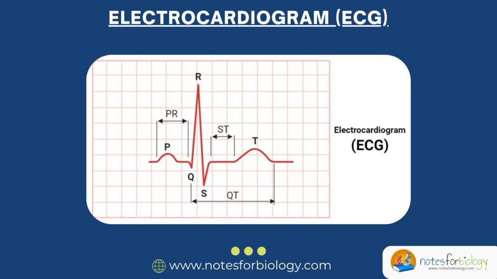
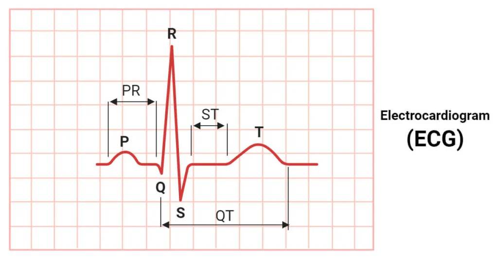
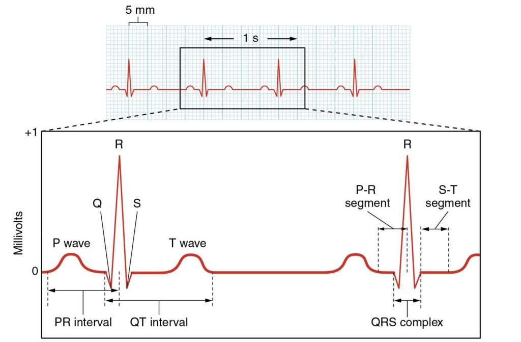
Electrocardiogram (ECG) Waves and Intervals
Each ECG tracing includes characteristic waves that represent specific events in the heart’s electrical cycle:
1. P Wave
- Atrial depolarization (contraction)
- Small upward deflection
2. QRS Complex
- Ventricular depolarization (contraction)
- Consists of:
- Q wave: Small downward deflection
- R wave: Sharp upward spike
- S wave: Downward deflection after R wave
3. T Wave
- Ventricular repolarization (recovery)
- Broad upward curve
4. U Wave (sometimes present)
Uncertain origin, may represent Purkinje repolarization
5. PR Interval
Time from start of atrial contraction to start of ventricular contraction
6. QT Interval
Time for ventricles to contract and then recover
Abnormalities in these waves or intervals can indicate a range of heart problems, such as arrhythmias, myocardial infarction, electrolyte imbalances, or conduction blocks.
Procedure of Electrocardiogram (ECG) Recording
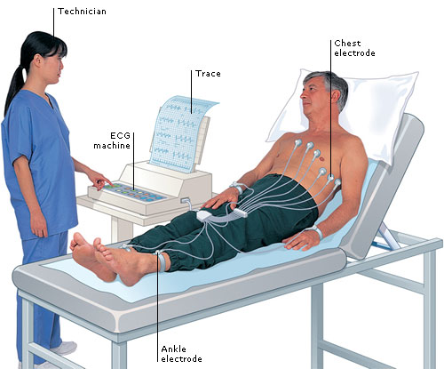
1. Preparation
- The patient lies down comfortably.
- Skin areas where electrodes will be placed are cleaned.
- Shaving may be done for better contact.
2. Electrode Placement
- Typically, 10 electrodes are placed:
- 6 on the chest (precordial leads)
- 1 on each limb (right arm, left arm, right leg, left leg)
3. Lead Configuration
- The 10 electrodes produce 12 leads:
- Limb leads: I, II, III, aVR, aVL, aVF
- Chest leads: V1 to V6
4. Recording the ECG
- The machine is switched on.
- Electrical activity is recorded for several seconds.
- Tracings are either printed or saved digitally.
5. Interpretation
- A healthcare provider interprets the waveform to identify abnormalities.
- Measurements like heart rate, rhythm, intervals, and wave heights are analyzed.
Types of Electrocardiogram (ECG)
Depending on the purpose and duration of monitoring, there are various types of ECGs:
1. Resting ECG
- Most common type
- Done while the patient is lying still
- Used for general cardiac assessment
2. Stress ECG (Exercise ECG)
- Done during physical exercise (like treadmill or bike)
- Evaluates heart function under stress
- Helpful in diagnosing ischemia or exercise-induced arrhythmias
3. Holter Monitoring
- Continuous ECG recording over 24 to 48 hours
- Used to detect intermittent arrhythmias or evaluate heart rhythm during daily activities
4. Event Recorder
- Worn for weeks
- Activated by the patient during symptoms
- Used to capture infrequent abnormal rhythms
5. Implantable Loop Recorder
- Inserted under the skin for long-term monitoring
- Detects unexplained fainting or irregular heartbeats
6. Telemetry Monitoring
- Real-time monitoring in hospitals
- Sends heart data to central monitoring systems
7. Fetal ECG
- Monitors fetal heart rate and rhythm during pregnancy
- Uses abdominal electrodes on the mother
Clinical Applications of ECG
1. Diagnosing Heart Disorders
- Myocardial infarction (heart attack)
- Angina
- Arrhythmias
- Pericarditis
- Cardiomyopathy
2. Monitoring Heart Health
- After surgery
- During medication therapy (e.g., antiarrhythmics)
- While using pacemakers or defibrillators
3. Pre-Surgical Evaluation
Routine ECGs before anesthesia
4. Emergency Medicine
Quick assessment of chest pain
5. Screening Athletes and Elderly
Identifies underlying heart risks
Advantages of ECG
- Non-invasive and painless
- Quick and easy to perform
- Cost-effective diagnostic tool
- Detects a wide range of cardiac problems
- No radiation involved
Limitations of ECG
- Provides only a snapshot in time
- May miss intermittent issues
- Interpretation requires trained personnel
- Can be affected by movement, electrode misplacement, or skin conditions
Safety Considerations
- ECG is very safe
- No electricity enters the body
- Only electrodes detect existing electrical activity
Conclusion
An Electrocardiogram (ECG) is a fundamental tool in cardiology that helps visualize the heart’s electrical system. By interpreting the characteristic waves and intervals on the ECG tracing, doctors can diagnose and monitor a wide range of heart conditions. Whether it is a simple check-up, a stress test, or a 24-hour Holter recording, the ECG remains one of the most accessible and informative tests in modern medicine.
Understanding how it works and what it reveals about our heart can empower patients and healthcare providers to make better decisions about heart health. With technology advancing, digital ECGs and portable devices are making it even easier to monitor the heart anytime, anywhere.
Three Key Summary
- ECG is a safe and essential test that records the heart’s electrical activity using skin electrodes.
- It is useful for detecting rhythm disorders, heart attacks, and many cardiac conditions.
- Various types of ECGs exist for different monitoring needs, from resting ECGs to long-term wearable monitors.
Frequently Asked Questions (FAQs)
Is ECG painful?
No, ECG is painless. It involves placing electrodes on the skin without any needles or shocks.
How long does a standard ECG take?
A typical resting ECG takes about 5 to 10 minutes.
Can ECG detect a heart attack?
Yes, ECG can show signs of current or past heart attacks, especially through changes in the ST segment or Q waves.
Related Articles