Introduction
The world of microorganisms, invisible to the naked eye, has fascinated scientists for centuries. Observing these tiny entities requires specialized tools—and among these, the darkfield microscope stands out for its ability to make transparent and unstained specimens come alive in glowing detail. Whether it’s viewing living bacteria, spirochetes, or minute aquatic organisms, darkfield microscopy offers a unique visual experience where objects appear brightly lit against a dark background.
This document provides a comprehensive and humanized overview of the darkfield microscope, including its definition, working principle, structure, applications, advantages, disadvantages, and a labeled diagram. The goal is to make this knowledge accessible and meaningful to students, professionals, and anyone curious about microbiological observation.
Table of Contents
1. What is a Darkfield Microscope?
Definition
A darkfield microscope is a type of optical microscope that uses a special illumination technique to make unstained and transparent specimens visible by scattering light so that only the light reflected or refracted by the specimen enters the objective lens. The specimen appears bright against a dark background—hence the name “darkfield.”
Key Characteristics:
- Uses oblique illumination
- Only scattered light is collected
- Background appears black; specimen appears glowing or bright
- Commonly used for live and unstained specimens
2. Principle of Darkfield Microscopy
The darkfield microscope works on the principle of light scattering.
How It Works:
- A special condenser blocks the central beam of light and allows only light rays that are angled.
- These angled rays pass around the specimen and do not enter the objective unless they are scattered.
- When the specimen deflects or refracts the angled light, it is scattered into the objective lens.
- The result: the background remains dark, while the specimen appears brightly illuminated.
Scientific Explanation:
- Unlike brightfield microscopy where light passes through the specimen, darkfield relies on Rayleigh scattering or Mie scattering to detect structures.
- It enhances contrast in specimens that are otherwise invisible or difficult to observe under normal light.
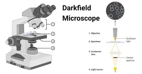
3. Components of a Darkfield Microscope
A darkfield microscope is based on a compound light microscope but includes some specialized components for darkfield imaging.
Main Components:
- Light Source
- Provides the illumination needed to view the specimen.
- Usually placed beneath the stage.
- Darkfield Condenser
- The heart of the system.
- Has a central opaque disc to block direct light and create a hollow cone of light.
- Stage
- Flat platform where the specimen slide is placed.
- Includes clips or mechanical stage to move the slide.
- Objective Lenses
- Magnify the image.
- High-quality objectives (preferably oil immersion for high magnification) are used.
- Eyepiece (Ocular Lens)
- Used to view the image formed by the objective lens.
- Usually 10x or 15x magnification.
- Adjustable Diaphragm and Mirror (for mirror-based microscopes)
- Controls light intensity and direction.
- Mechanical Arm and Base
- Support and hold the microscope structure.
4. Types of Darkfield Microscope
A. Simple Darkfield Microscopy
- Uses a simple darkfield condenser.
- Suitable for low to medium magnification.
B. Oil Immersion Darkfield Microscopy
- High-magnification darkfield imaging.
- Requires a special oil condenser and immersion oil for objectives.
- Commonly used to view bacteria and spirochetes.
C. Reflective Darkfield Microscopy (Used in Metallurgy)
- Used for opaque objects like metals.
- Illumination comes from above.
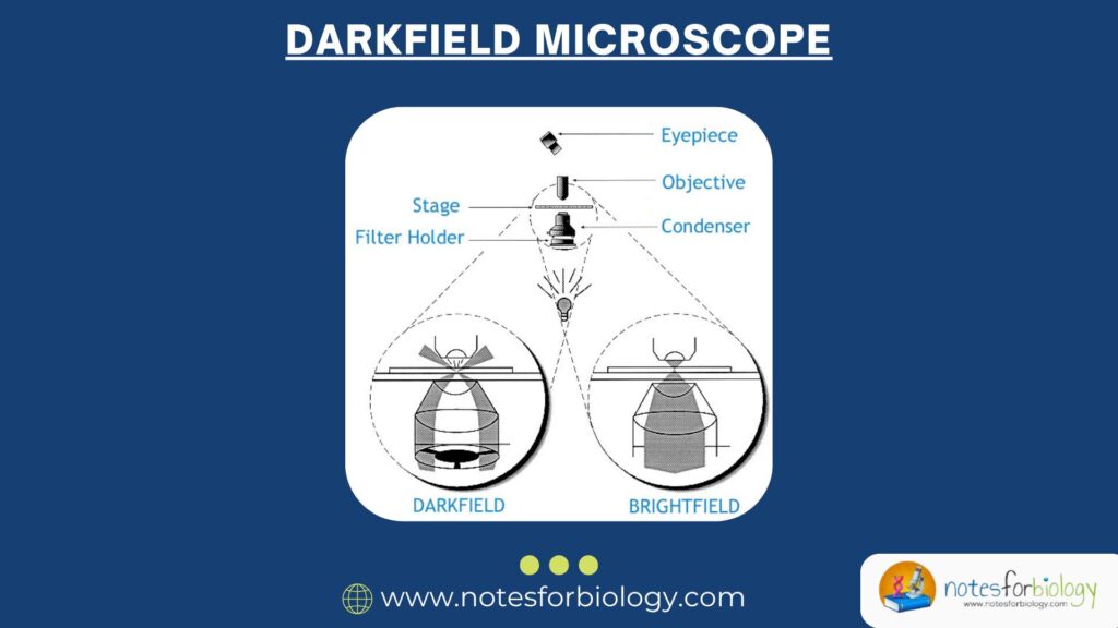
5. Diagram of a Darkfield Microscope
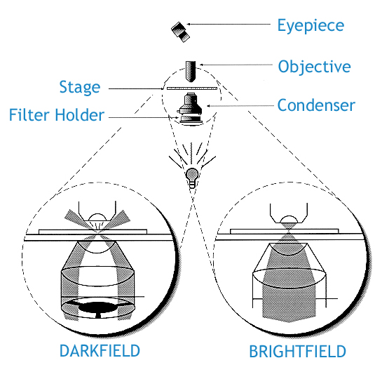
This simplified diagram shows how light is manipulated below the specimen to create a darkfield effect.
6. Preparation of Specimen for Darkfield Microscopy
- Do not stain the specimen; most dyes absorb or scatter light in ways incompatible with darkfield imaging.
- Use coverslips to flatten the specimen and prevent drying.
- Keep slides clean and dry to avoid scattering from dust or water.
- Live specimens should be placed in a drop of water.
7. Applications and Uses of Darkfield Microscopy
A. Medical Microbiology
- Detection of spirochetes, e.g., Treponema pallidum (syphilis-causing bacterium)
- Borrelia burgdorferi (causes Lyme disease)
B. Parasitology
Visualize protozoans and small parasitic larvae
C. Aquatic Biology
Study of plankton, algae, and small aquatic organisms
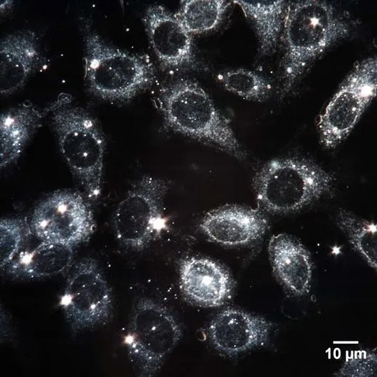
D. Cell Biology
Observe live cells, especially those with transparent structures
E. Industrial Uses
Examine surface flaws in metals and materials
F. Teaching and Research
- Engages students with visually striking images
- Used in advanced microscopy training
8. Advantages of Darkfield Microscopy
- Enhanced Contrast
- Makes invisible, unstained structures visible
- Live Specimen Observation
- No need to kill or stain organisms
- Simple to Use
- Modifies basic microscopes with minimal adjustments
- Non-Invasive
- Preserves natural state of specimen
- Cost-Effective
- Does not require expensive stains or dyes
9. Disadvantages and Limitations
- Limited to Thin Specimens
- Thick specimens scatter too much light and blur the image
- Low Light Intensity
- May require strong light sources or sensitive detectors
- Artifacts and Debris Can Appear Bright
- Dirt or dust may be mistaken for real structures
- Low Resolution Compared to Other Techniques
- Cannot compete with phase-contrast or electron microscopy in detail
- Difficult to Photograph or Record
- Requires special cameras or exposure settings
10. Comparison: Darkfield vs Brightfield Microscopy
| Feature | Darkfield | Brightfield |
|---|---|---|
| Background | Dark | Bright/White |
| Specimen Appearance | Bright and glowing | Dark or stained |
| Contrast | High (natural) | Often low (requires stains) |
| Live Cell Observation | Excellent | Limited |
| Complexity | Requires darkfield condenser | Simple setup |
11. Tips for Effective Darkfield Microscopy
- Keep optics clean: Dust can distort light scattering.
- Align condenser carefully: Misalignment reduces contrast.
- Use coverslips to flatten liquid specimens.
- Start with low magnification, then increase gradually.
- Adjust diaphragm and light intensity to fine-tune visibility.
12. Innovations in Darkfield Microscopy
- Digital darkfield microscopy: Combines traditional technique with digital cameras and image processing.
- Darkfield fluorescence microscopy: Merges darkfield with fluorescent markers.
- High-resolution adaptations: Incorporate advanced optics for improved detail.
Conclusion
Darkfield microscopy is a powerful and accessible technique that allows us to explore the invisible world of living cells, microorganisms, and minute particles with dazzling clarity. By illuminating only the light scattered by the specimen, it provides dramatic contrast and makes delicate structures visible without staining or destroying the sample.
While it may not offer the resolution of advanced techniques like electron microscopy, darkfield microscopy remains a vital tool in microbiology, medical diagnostics, and biological research. For students and scientists alike, it opens a glowing window into the microscopic universe—illuminating the unseen, one bright speck at a time.
FREQUENTLY ASKED QUESTIONS
Can I use darkfield microscopy to see viruses?
No, viruses are too small to be seen with light microscopy, including darkfield. Electron microscopy is needed.
Why is the specimen glowing in darkfield microscopy?
Because only scattered light from the specimen enters the objective, making it appear bright against a dark background.
Do I need to stain my samples?
No, in fact, staining is discouraged in darkfield microscopy as it interferes with light scattering.
Related Articles