The centrosomes is a key cellular structure that plays a crucial role in organizing microtubules and ensuring proper cell division in eukaryotic cells. Composed of a pair of centrioles and surrounding pericentriolar material (PCM), the centrosome acts as the primary microtubule-organizing center (MTOC) and helps maintain the structural integrity and polarity of the cell. This comprehensive overview discusses the definition, detailed structure, functions, associated proteins, clinical relevance, and recent research insights into the centrosome.
1. Definition of the Centrosome
The centrosome is an organelle located near the nucleus of animal cells. It is the principal microtubule-organizing center and serves as a hub for microtubule nucleation, anchoring, and organization. The centrosome consists of two perpendicular centrioles surrounded by amorphous, electron-dense pericentriolar material (PCM). It plays a pivotal role in forming the mitotic spindle during cell division and is essential for cellular shape, polarity, and intracellular transport.
Summary of Centrosome
- The centrosome consists of two perpendicular centrioles (mother and daughter) surrounded by pericentriolar material.
- It plays a key role in organizing microtubules, aiding in cell division, intracellular transport, and maintaining cell shape.
- The centrosome helps form the mitotic spindle during cell division, ensuring proper chromosome separation.
Table of Contents
2. Structure of the Centrosomes
The centrosomes is made up of two main components:
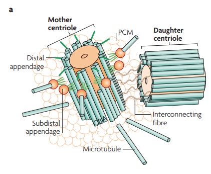
2.1 Centrioles
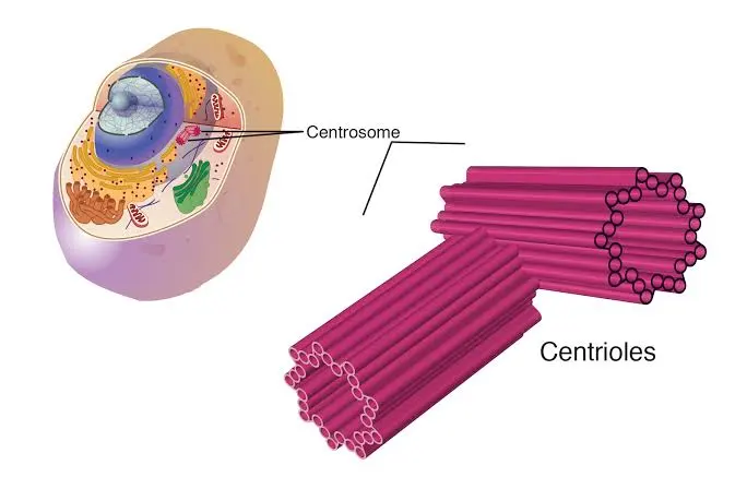
Each centrosomes contains a pair of centrioles. These are cylindrical structures, each measuring around 250 nm in diameter and 500 nm in length. Each centriole is composed of nine triplets of microtubules arranged in a radial pattern, known as the “9+0” configuration. The centrioles are arranged at right angles to each other and play an important role in initiating and organizing the mitotic spindle.
2.2 Pericentriolar Material (PCM)
Surrounding the centrioles is the PCM, an amorphous cloud of proteins that assists in microtubule nucleation and anchoring. The PCM contains important proteins such as γ-tubulin, pericentrin, and CDK5RAP2. These proteins provide the structural framework and nucleating sites for microtubules.
2.3 Centrosome Duplication
The centrosome duplicates once per cell cycle, typically during the S phase. Each new centrosome inherits one of the original centrioles and forms a daughter centriole adjacent to it. This process is tightly regulated to ensure that cells maintain only two centrosomes, preventing abnormal cell division and chromosomal missegregation.
3. Functions of the Centrosome
The centrosomes serves multiple vital roles in eukaryotic cells:
3.1 Microtubule Organization
The centrosomes acts as the main site for microtubule nucleation. The γ-tubulin ring complexes (γ-TuRCs) within the PCM initiate the polymerization of tubulin dimers, forming the microtubule cytoskeleton. These microtubules are involved in maintaining the cell’s shape, facilitating intracellular transport, and segregating chromosomes during cell division.
3.2 Mitotic Spindle Formation
During mitosis, centrosomes duplicate and migrate to opposite poles of the cell, organizing the bipolar mitotic spindle. This structure is critical for the accurate segregation of chromosomes into daughter cells. Proper centrosome function ensures genomic stability, while centrosome abnormalities can lead to aneuploidy and cancer.
3.3 Establishing Cell Polarity
In polarized cells, such as epithelial cells and neurons, the centrosome helps organize the microtubule network to maintain cell polarity and directional movement. It is involved in positioning the nucleus, Golgi apparatus, and other organelles.
3.4 Ciliogenesis
One of the centrioles in the centrosomes, known as the mother centriole, can migrate to the cell membrane and convert into a basal body. This basal body then initiates the formation of cilia and flagella hair-like structures involved in cell motility and signaling.
4. Associated Proteins and Regulatory Molecules
The centrosome contains many essential proteins that contribute to its function and duplication. These include:
- γ-tubulin: Nucleates microtubules from the PCM.
- Pericentrin: Provides structural support and anchors other PCM proteins.
- CDK5RAP2: Regulates microtubule nucleation.
- PLK4: A kinase that initiates centriole duplication.
- CEP192, CEP152, STIL, SAS-6: Involved in the assembly and elongation of centrioles.
These proteins form complex networks that regulate the formation, function, and maintenance of the centrosome.
5. Clinical Significance and Disease Associations
Abnormalities in centrosomes number or function are linked to several diseases:
5.1 Cancer
Supernumerary centrosomes are often found in cancer cells. These extra centrosomes can form multipolar spindles, leading to chromosomal missegregation and genomic instability. Mutations in genes regulating centrosome duplication (e.g., PLK4, CEP192) are associated with tumor progression.
5.2 Microcephaly
Defects in centrosomal proteins can impair neural progenitor cell division during brain development, resulting in microcephaly (a condition characterized by reduced brain size). Mutations in genes like ASPM, CDK5RAP2, and CEP135 are commonly implicated.
5.3 Ciliopathies
As the mother centriole gives rise to basal bodies and cilia, centrosomal defects can disrupt ciliogenesis. This leads to a group of disorders called ciliopathies, which include:
- Polycystic Kidney Disease (PKD)
- Bardet-Biedl Syndrome (BBS)
- Meckel-Gruber Syndrome (MKS)
- Joubert Syndrome
These diseases typically affect organs like the kidneys, liver, and brain and can cause symptoms ranging from vision loss to developmental delay.
6. Technological and Research Advances
Recent advances in science and technology have greatly enhanced our ability to study the centrosomes in detail. Four key technologies used in centrosome research are:
6.1 Super-Resolution Microscopy
This advanced form of light microscopy overcomes the diffraction limit, allowing scientists to observe cellular structures at the nanometer scale. It is particularly useful for visualizing the precise arrangement of proteins in the centrosome.
6.2 Cryo-Electron Tomography (Cryo-ET)
Cryo-ET is a powerful imaging technique that combines cryogenic freezing and electron microscopy to produce high-resolution 3D images of cellular structures in their near-native state. It helps scientists understand the ultrastructure of centrioles and PCM.
6.3 Live-Cell Imaging
This technique allows researchers to observe living cells over time using fluorescent markers. It helps track centrosome dynamics, such as duplication, migration, and mitotic spindle formation in real-time.
6.4 Proteomics and Gene Editing (e.g., CRISPR-Cas9)
Proteomics is the large-scale study of proteins and their interactions, enabling researchers to identify new centrosomal components. Gene editing tools like CRISPR-Cas9 allow precise manipulation of genes to study the function of centrosomal proteins and their impact on cell behavior.
These tools are revolutionizing centrosome biology by uncovering new functions, interactions, and disease links, and paving the way for targeted therapies.
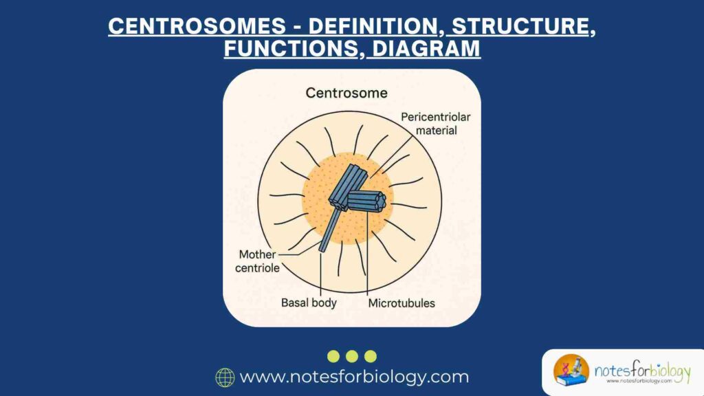
7. Diagram of the Centrosomes
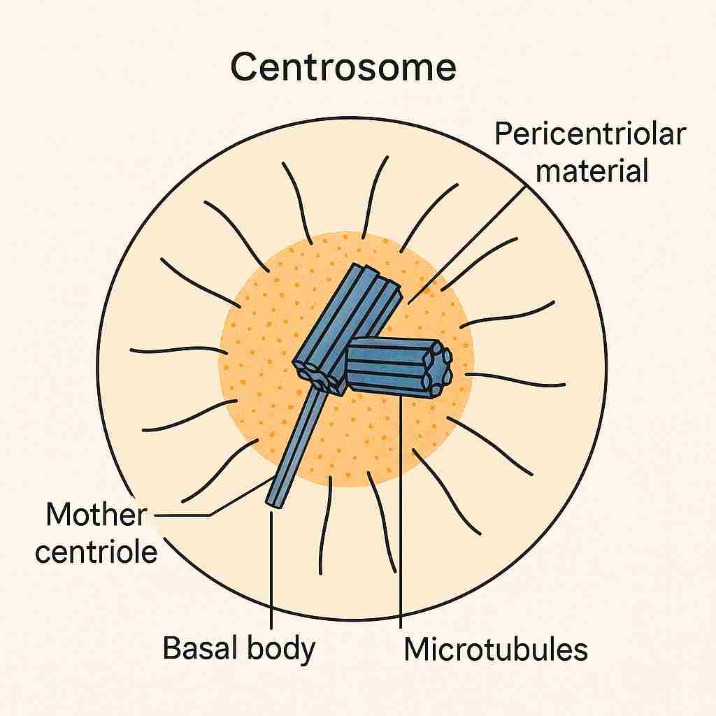
A simple diagram of the centrosomes typically includes:
- Two perpendicular centrioles (mother and daughter)
- Pericentriolar material (surrounding the centrioles)
- Microtubules radiating outward
- Basal body formed from the mother centriole (if showing ciliogenesis)
This visual representation helps illustrate how the centrosome serves as a hub for microtubule organization and cellular coordination.
8. Conclusion
The centrosome is a fundamental organelle in animal cells, responsible for organizing the microtubule network, regulating cell division, establishing polarity, and facilitating the formation of cilia. Its structural complexity and functional versatility highlight its importance in normal cellular physiology and development. Dysfunction of the centrosome can lead to a range of human diseases, underscoring the need for continued research. As we deepen our understanding of centrosome biology, we unlock new opportunities for diagnosing and treating conditions related to its dysfunction.
Frequently Asked Questions (FAQs)
What is a centrosome?
A centrosome is a small organelle found in animal cells that acts as the main microtubule-organizing center (MTOC). It consists of two centrioles surrounded by a protein-rich material called pericentriolar material (PCM). Centrosomes play a key role in cell division, maintaining cell shape, and organizing microtubules for intracellular transport.
What is the structure of a centrosome?
A centrosome has two barrel-shaped centrioles (mother and daughter) arranged at right angles to each other. The mother centriole has appendages that help anchor microtubules, while the pericentriolar material (PCM) contains proteins like γ-tubulin, which helps in microtubule formation.
What are the functions of centrosomes?
Centrosomes help in cell division by forming the mitotic spindle, which separates chromosomes. They also organize microtubules to maintain cell structure and assist in cilia and flagella formation (hair-like structures for movement). Without centrosomes, cells may divide incorrectly, leading to diseases like cancer.
Related Articles