Introduction
Centrioles are cylindrical, tube-like structures found in most eukaryotic cells. Cells are the basic building blocks of life, and each part of a cell plays a unique and crucial role in ensuring the cell functions properly. Among the many organelles in a cell, centrioles are small but very important structures that often go unnoticed. Despite their tiny size, centrioles play a major role in cell division, organization of microtubules, and formation of structures like cilia and flagella. Understanding centrioles is vital for grasping how cells reproduce and maintain their internal structure.
Table of Contents
Definition of Centrioles
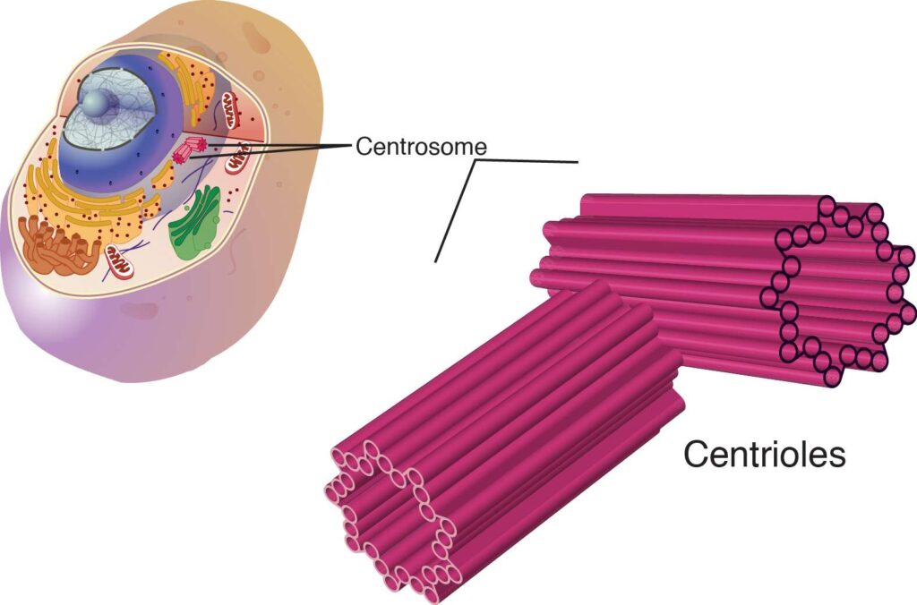
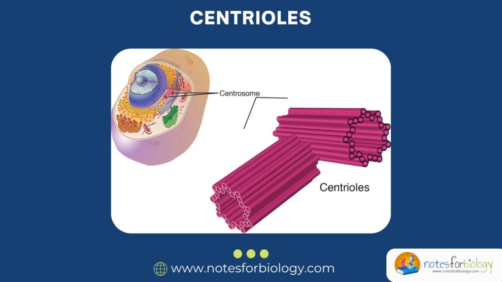
Centrioles are cylindrical, tube-like structures found in most eukaryotic cells. They are part of a larger structure known as the centrosome, which acts as the main microtubule organizing center (MTOC) of the cell. A typical centriole is composed of microtubules arranged in a specific pattern and is usually found in pairs.
To define it simply: Centrioles are cylindrical structures made up of microtubules that help in the organization of spindle fibers during cell division and contribute to the formation of cilia and flagella.
They are especially prominent in animal cells, although similar structures are sometimes seen in lower plants. Higher plants usually lack centrioles but still manage cell division through other means.
Location in the cell
he Cell
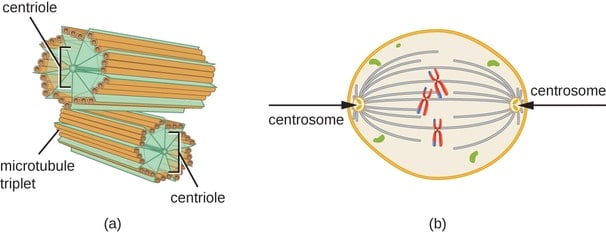
Centrioles are usually located near the nucleus of animal cells. Two centrioles are typically arranged perpendicular to each other, forming a structure known as the centrosome. During the process of cell division, this centrosome plays a vital role in organizing the mitotic spindle, which separates chromosomes into daughter cells.
Structure of Centrioles
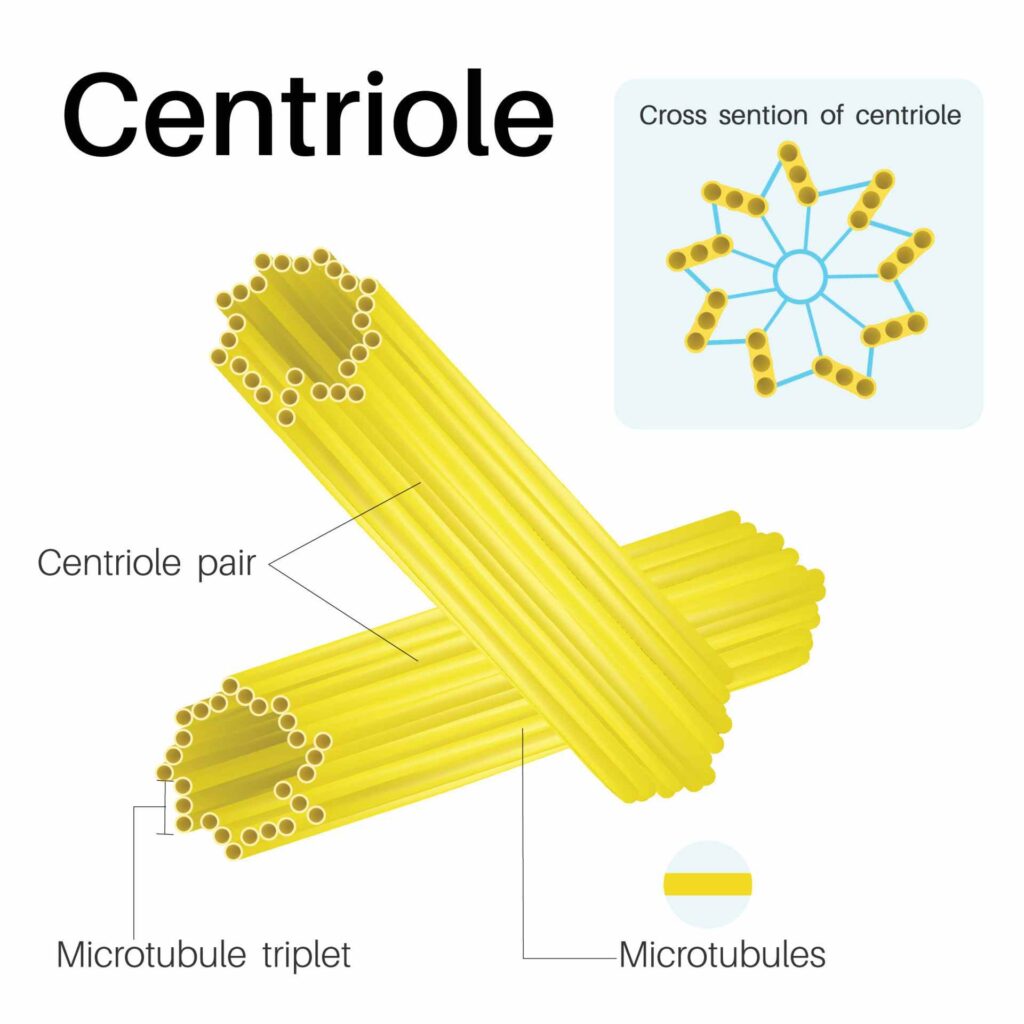
The structure of a centriole is both simple and elegant. Each centriole is about 300–500 nanometers long and 200–250 nanometers in diameter. Here’s a breakdown of its structure:
- Microtubule Arrangement:
- Centrioles are made up of microtubules, which are cylindrical tubes composed of tubulin proteins.
- Each centriole contains nine triplets of microtubules arranged in a circular pattern (often referred to as a “9+0” pattern).
- Each triplet consists of three microtubules: A, B, and C.
- Microtubule A is a complete microtubule, while B and C share walls with the adjacent microtubules.
- Pericentriolar Material (PCM):
- The centrioles are surrounded by an amorphous, protein-rich matrix called the pericentriolar material.
- PCM plays a role in anchoring microtubules and regulating their growth.
- Linking Fibers:
- The two centrioles in a centrosome are connected by protein fibers, which help maintain their orientation.
- No Membrane:
- Unlike many other organelles, centrioles are non-membranous structures.
Diagram of a Centriole
Functions of Centrioles
Though small, centrioles have several important functions in the cell:
Cell Division:
- During mitosis and meiosis, centrioles help in organizing the spindle fibers that separate chromosomes.
- The centrosome duplicates before cell division, and each pair migrates to opposite poles of the cell.
- The spindle fibers extend from the centrioles and attach to chromosomes to pull them apart.
Microtubule Organization:
- Centrioles organize and anchor the microtubules of the cell’s cytoskeleton.
- This helps maintain cell shape and aids in intracellular transport.
Formation of Cilia and Flagella:
- Centrioles serve as basal bodies from which cilia and flagella are formed.
- These structures are important for cell movement and fluid movement across the cell surface.
Establishing Cell Polarity:
- The position of centrioles helps determine the spatial arrangement of cell components.
- This is crucial during development and for processes like asymmetric cell division.
Role in Development and Differentiation:
Centrioles are involved in signaling pathways that regulate stem cell behavior, differentiation, and development.
Centrosome vs Centriole
It’s important to distinguish between centrioles and the centrosome:
- A centriole is a single cylindrical structure made up of microtubules.
- A centrosome is a complex that includes a pair of centrioles and the surrounding pericentriolar material.
- The centrosome acts as the MTOC, organizing the cell’s microtubules, especially during cell division.
Centrioles in Different Organisms
- Animal Cells: Almost all animal cells contain a pair of centrioles.
- Plant Cells: Higher plants typically do not have centrioles. Instead, they use other structures for microtubule organization.
- Fungi and Protists: Some species possess centrioles, especially those with flagella.
This indicates that centrioles are not universally present in all eukaryotes, but they are essential where they are found.
Abnormalities Related to Centrioles
Problems with centrioles can lead to serious cellular and developmental disorders:
- Cancer:
- Abnormal duplication of centrioles can result in defective mitotic spindles.
- This may lead to aneuploidy (abnormal number of chromosomes), a hallmark of many cancers.
- Ciliopathies:
- Defects in basal bodies (modified centrioles) can lead to diseases related to cilia, such as respiratory issues and polycystic kidney disease.
- Neurodevelopmental Disorders:
- Faulty centrioles can disrupt brain development and cell differentiation.
Recent Research and Discoveries
Modern cell biology has revealed many fascinating aspects of centrioles:
- Self-replication: Centrioles duplicate once per cell cycle, typically during the S-phase, through a highly regulated process.
- Centriole Biogenesis: This involves a sequence of steps starting with the formation of a small structure called a “procentriole,” which elongates and matures over time.
- Proteins Involved: Proteins like SAS-6, STIL, and PLK4 are essential for centriole duplication and stability.
- Super-resolution Microscopy: This has allowed scientists to visualize centriole structure in unprecedented detail, enhancing our understanding of their role.
Key Points to Remember
- Centrioles are microtubule-based cylindrical structures found mainly in animal cells.
- Each centriole is made of 9 triplets of microtubules.
- A pair of centrioles, along with pericentriolar material, makes up the centrosome.
- Their main functions include cell division, formation of cilia/flagella, and microtubule organization.
- Defects in centrioles can lead to diseases like cancer and genetic disorders.
- Plant cells do not usually contain centrioles, showing alternative pathways of microtubule organization.
Conclusion
Centrioles, though small and often overlooked, are essential for many cellular processes. Their role in cell division ensures proper distribution of genetic material, which is crucial for growth and development. They also help cells maintain their shape and create important motile structures like cilia and flagella. Understanding the structure and function of centrioles provides insight into the intricate machinery that drives life at the cellular level. As science progresses, new discoveries about these humble organelles continue to shed light on their significance in health and disease.
Their complexity, precision, and multifaceted functions make centrioles a perfect example of the marvels hidden inside every living cell.
FREQUENTLY ASKED QUESTIONS
What are centrioles?
Centrioles are cylindrical structures made up of microtubules, found mostly in animal cells. They are essential for organizing spindle fibers during cell division and for the formation of cilia and flagella.
Where are centrioles located in the cell?
Centrioles are usually found near the nucleus in animal cells, within a region called the centrosome. They typically exist in pairs positioned perpendicular to each other.
What is the difference between a centriole and a centrosome?
A centriole is a single microtubule-based structure, while a centrosome is a complex that includes a pair of centrioles and surrounding pericentriolar material (PCM). The centrosome acts as the cell’s microtubule-organizing center (MTOC).
Related Articles