
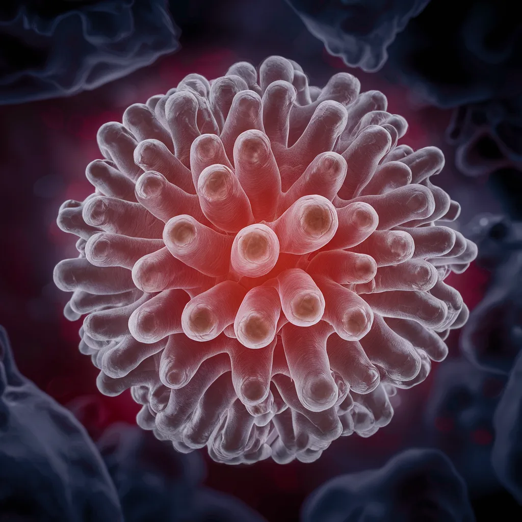
Klebsiella granulomatis, formerly Calymmatobacterium granulomatis, is a Gram-negative bacteria that causes the sexually transmitted illness granuloma inguinale (donovanosis). Biochemical testing is critical for identifying and distinguishing Klebsiella granulomatous from other Klebsiella species and similar diseases.
Table of Contents
What is Klebsiella granulomatis?
Klebsiella granulomatis is a Gram-negative rod-shaped bacteria that causes granuloma inguinale (donovanosis), a sexually transmitted infection. This bacterium primarily affects the vaginal and inguinal regions, resulting in persistent ulcers and granulomatous inflammation. Biochemical testing is critical for identifying and distinguishing Klebsiella granulomatous from other Klebsiella species and similar diseases.
Here’s a detailed explanation of the biochemical tests used to identify Klebsiella granulomatis:
1. Gram Stain

Principle:
The Gram stain is a differential staining technique that uses the makeup of bacteria’s cell walls to classify them as Gram-positive or Gram-negative.
Procedure:
Make a bacterial stain on a glass slide and heat-fix it.
Stain with crystal violet for one minute.
Rinse with water and apply the iodine solution for 1 minute.
Decolorize for 10-20 seconds with alcohol or acetone.
Counterstain with safranin for 1 minute, then rinse with water.
Result:
Gram-negative rods appear pink/red because of their thin peptidoglycan coating, which does not preserve the crystal violet-iodine complex after decolorization.
2. Oxidase Test
Principle:
This test determines the presence of cytochrome c oxidase, an enzyme of the bacterial electron transport chain.
Procedure:
Using a sterile loop or swab, collect a small amount of bacterial culture. Apply the culture to a piece of filter paper impregnated with oxidase reagent (tetramethyl-p-phenylenediamine).
Result:
Negative: No color change indicates the absence of cytochrome c oxidase
3. Catalase Test
Principle:
The catalase test determines the presence of catalase, an enzyme that converts hydrogen peroxide into water and oxygen.
Procedure:
Place a drop of hydrogen peroxide (H2O2) onto a glass slide.
Using a sterile loop, add a small amount of bacterial culture to the drop of hydrogen peroxide.
Result:
Positive: Bubbling or effervescence signals the release of oxygen gas from the decomposition of hydrogen peroxide.
4. Urease Test
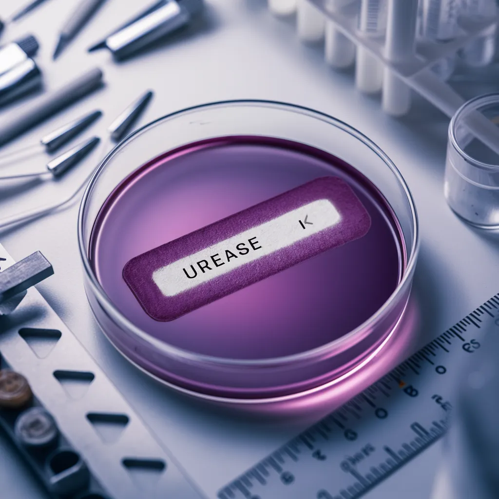
Principle:
This test identifies urease, an enzyme that converts urea to ammonia and carbon dioxide, raising the pH of the medium.
Procedure:
Inoculate the bacterial culture into a medium containing urea and a pH indicator (such as phenol red).
Incubate at 37°C for 24 to 48 hours.
Result:
Positive: A change in hue to pink/red indicates the formation of ammonia, which raises the pH of the medium.
5. Indole Test
Principle:
The indole test measures bacteria’s capacity to make indole from the amino acid tryptophan via the enzyme tryptophanase.
Procedure:
Incubate the bacterial culture in tryptone broth at 37°C for 24–48 hours.
Add a few of drops of Kovac’s reagent to the culture.
Result:
Negative: No color change (yellow) shows the lack of indole formation.
6. Citrate Utilization Test

Principle:
This test checks if a bacteria can use citrate as its sole carbon source and ammonium ions as its only nitrogen source.
Procedure:
Incubate the Simmon’s citrate agar slant with the bacterial culture.
Incubate at 37°C for 24 to 48 hours.
Result:
Positive: A blue color change suggests citrate consumption, which results in medium alkalinization.
7. Triple Sugar Iron (TSI) Agar
Principle:
TSI agar distinguishes bacteria based on their capacity to ferment glucose, lactose, and sucrose and create hydrogen sulfide (H2S).
Procedure:
Stab the buttocks and streak the bacterial culture onto the slope of the TSI agar.
Incubate at 37°C for 18 to 24 hours.
Result:
Acid/Acid (A/A), Gas Production, and No H2S: The yellow slant and butt suggest that glucose, lactose, and/or sucrose have been fermented. Gas bubbles and fissures in the agar indicate gas production. The absence of black precipitate suggests no H2S generation.
8. Methyl Red (MR) Test
Principle:
The MR test detects the formation of stable acids during glucose fermentation, indicating mixed acid fermentation.
Procedure:
Inoculate MR-VP broth with the bacterial cultures.
Incubate at 37°C for 24 to 48 hours.
Add a few drops of the methyl red indicator to the culture.
Result:
Negative: No color change (yellow) indicates the absence of steady acid formation during glucose fermentation.
Frequently Asked Question(FAQ)
What is Klebsiella granulomatis?
Klebsiella granulomatis, formerly Calymmatobacterium granulomatis, is a Gram-negative bacteria that causes the sexually transmitted illness granuloma inguinale (donovanosis). Biochemical testing is critical for identifying and distinguishing Klebsiella granulomatous from other Klebsiella species and similar diseases.
What are the biochemical tests used to identify Klebsiella granulomatis?
The biochemical tests used to identify Klebsiella granulomatis are:
1.Gram Stain
2. Oxidase Test
3. Catalase Test
4 .Urease Test
5. Citrate Utilization Test
6. Indole Test
Related Articles