
The main application of auramine-rhodamine staining, a fluorescent staining method, is the identification of acid-fast bacteria, particularly Mycobacterium species like Mycobacterium tuberculosis. This technique takes advantage of specific dyes’ characteristics to attach to the mycolic acid in acid-fast organisms’ cell walls and cause them to fluoresce when viewed using a fluorescence microscope.
Table of Contents
An age of improved staining techniques that are quick, more adaptable, and dependable in result interpretation has emerged as a result of the advancement of staining approaches. This is one of the methods that was adapted from the Acid Fast staining method, also referred to as the Ziehl-Neelsen Staining of Mycobacterium spp. The type of chemicals employed for staining and the method of observation are the main distinctions between these two approaches.
The Truant auramine-rhodamine stain, also known as the auramine-rhodamine staining technique, is a histology stain that is used to show the structure of the bacterial bacilli-cell and is also used to stain and demonstrate the presence of Acid Fast-bacilli under a fluorescent microscope.
Hagemann (1937) introduced the use of fluorescent dyes to increase the sensitivity of demonstrating Mycobacterial bacteria. Truant, Brett, and Thomas (1962) analyzed the significance of using fluorescent dyes for acid-fast bacilli and noted that there was a higher yield in demonstrating high numbers of positive acid-fast bacilli. In comparison to Ziehl Neelsen Staining procedures, this method performed better.
When auramine and rhodamine were combined, better outcomes were obtained than when the dyes were used separately. In addition, it was discovered that the complex moved more quickly than acid-fast staining.
Principle of Auramine-Rhodamine
The principle behind Auramine-Rhodamine staining involves the use of fluorescent dyes that specifically bind to the lipid-rich cell walls of acid-fast bacteria. When exposed to ultraviolet (UV) light or blue light, these dyes fluoresce, allowing for the visualization of the bacteria.
- Auramine O: An acid-fast organism-staining yellow dye.
- Improves contrast and fluorescence when combined with rhodamine B.
Procedure of Auramine-Rhodamine
- On a sterile, tiny glass slide, make a thin smear of the specimen and carefully heat fix it, taking care not to overheat.
- Make sure the smear is adequately stained by adding an adequate amount of Auramine-Rhodamine Dyes (Flooding) and letting it stand for 15 minutes. Avoid using heat.
- Rinse the stained smear with water until the effluent is colorless. Since chlorine obstructs fluorescence, make sure the water is free of chlorine, preferably through distillation.
- After adding the decolorizing agent (acid-alcohol) and letting it sit for two to three minutes to remove the stain, thoroughly wash the slide with distilled water, shaking off any extra water.
- For precisely two minutes, cover the streaks with potassium permanganate (counterstain). It should be noted that prolonged counterstaining can suppress the bacilli’s fluorescence.
- After giving it a good rinse with distilled water, let it air dry. Avoid blotting.
- Verify by looking under a fluorescent microscope with an oil immersion at 400X or a K530 excitation filter and BG12 or G-362 excitation filter and LP 420 barrier.
Interpretation
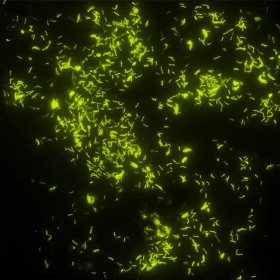
- Positive Outcome: The presence of acid-fast bacteria is indicated by bright yellow or orange-red fluorescent rods.
- Negative Outcome: The lack of fluorescent rods indicates that the sample does not contain any acid-fast bacteria.
Advantages
- More sensitive than the conventional Ziehl-Neelsen staining method.
- Speed: Quick identification of bacteria that grow quickly in acidic environments.
- Clarity: A distinct and contrasted image is produced via fluorescent staining.
Disadvantages
- Equipment: Fluorescence microscope is required; however, not all laboratories may have one.
- Specificity: Although sensitive, it might not be able to differentiate between several Mycobacterium species without additional research. These dyes, rhodamine and auramine, may cause cancer.
- Potassium permanganate with acid alcohol can irritate the respiratory system, eyes, and skin.
- The fluorescing organism’s brilliance may be lost if it is exposed to the counterstain excessively.
- It is important to observe stains within 24 hours of staining in order to prevent the fluorescence from fading.
Application of Auramine-Rhodamine
Diagnostic tool for tuberculosis: widely used to quickly identify Mycobacterium tuberculosis in clinical samples.
Research: Used in lab settings to examine the characteristics of acid-fast organisms.
An effective and sensitive technique for identifying acid-fast bacteria is aureamine-rhodamine staining, which is especially helpful in the diagnosis of tuberculosis. It is an effective tool for seeing these microorganisms in clinical and scientific settings since it uses fluorescent dyes.
Frequently Asked Question(FAQ)
What is the auramine stain method?
Make sure the smear is adequately stained by adding an adequate amount of Auramine-Rhodamine Dyes (Flooding) and letting it stand for 15 minutes. Avoid using heat. Rinse the stained smear with water until the effluent is colorless.
What is Auramine-Rhodamine used for?
Auramine colorants are employed as dye ingredients in ink ribbons, ballpoint pastes, oils and waxes, carbon paper, and leather, jute, and tanned cotton dyes.
What is the color of Auramine-Rhodamine Stain?
Auramine O, often known as Basic Yellow 2, is a fluorescent stain made of diarylmethane. Auramine O crystals that are yellow in color when pure.
How sensitive is Auramine-Rhodamine Stain?
An acid-fast stain’s estimated detection limit is 5,000–10,000 bacilli/mL of sputum; its overall sensitivity ranges from 22% to 78%.
Related Article