Ascaris lumbricoides is a species of parasitic roundworm (nematode) that infects the intestines of humans. It is one of the largest and most common human intestinal parasites. The adult worms reside in the lumen of the small intestine, where they can cause a range of health problems, from mild gastrointestinal discomfort to severe malnutrition and intestinal blockage. The lifecycle of Ascaris lumbricoides includes both larval and adult stages, involving migration through various body tissues before settling in the intestines. Transmission occurs through the ingestion of eggs present in contaminated soil, food, or water, typically in areas with poor sanitation.
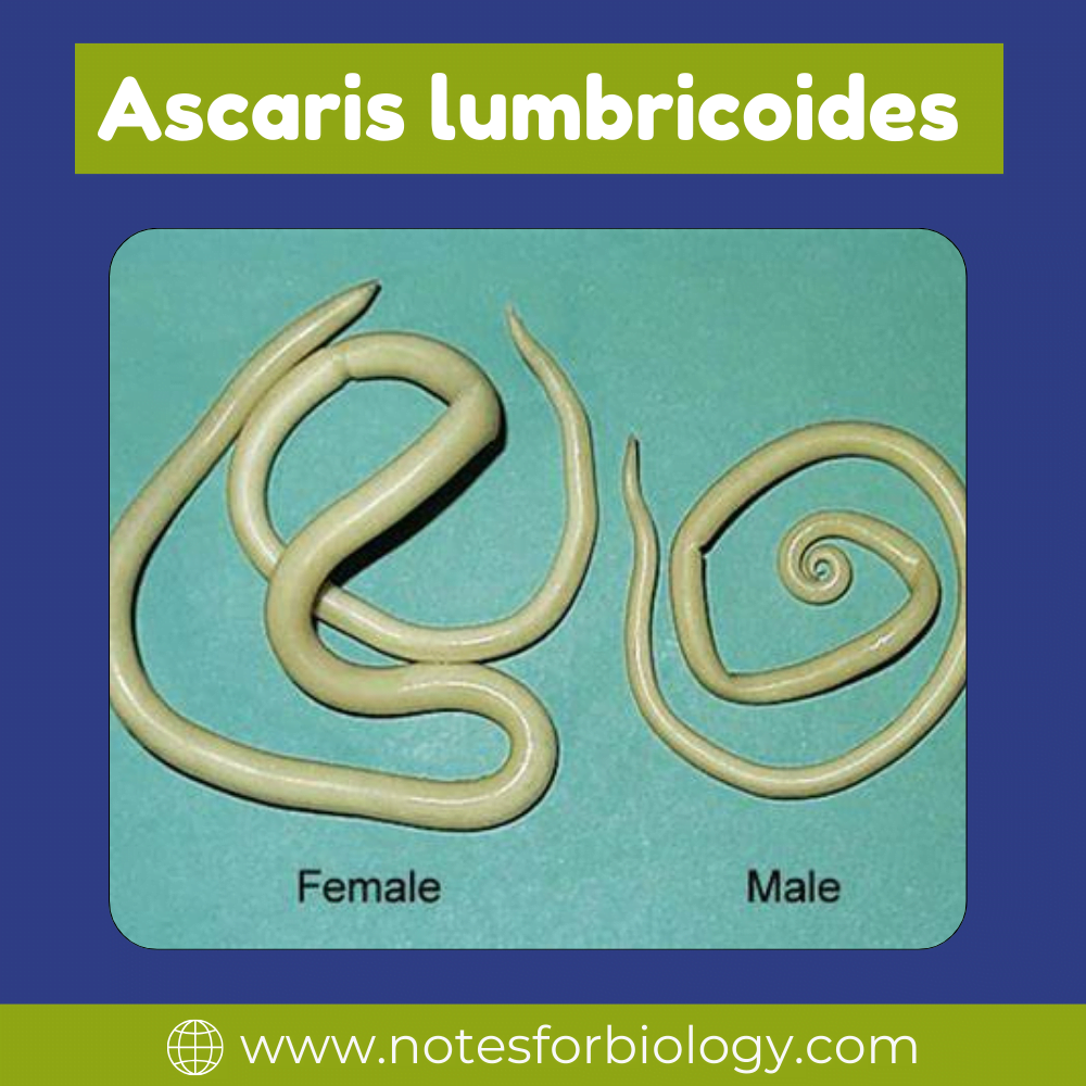
Table of Contents
Habitat of Ascaris lumbricoides
External Environment
Ascaris lumbricoides eggs exit the human body through excrement. They may persist in soil for long periods, especially in warm, wet circumstances. It is cosmopolitan in distribution.
Intermediate Hosts
When Ascaris eggs are consumed by suitable intermediate hosts such as earthworms or insects, they can develop but do not grow into adult worms. Instead, they act as carriers for eggs to reach the infective stage.
Human Host
The larvae hatch in the human small intestine after ingesting infected eggs from contaminated food, drink, or soil. The mature worms then live in the small intestine’s lumen, where they eat and breed, finishing their life cycle.
External Morphology of Ascaris lumbricoides
Shape and Size
It is cylindrical, long, and gradually tapered at both ends, with the front end being less thick than the back. Among nematodes, it is the largest. It exhibits dimorphism in a sexual sense. The genders are different. The female has a straight tail and measures 20 to 40 cm in length and 4-6 mm in diameter. The male has a ventrally coiled tail and is smaller, reaching between 15 and 31 cm in length and 2-4 mm in diameter.
Shades of colour
The nematodes are often colourless. And newly expelled worm is pinkish-yellow in appearance, eventually becoming white.
Cuticle
Ascaris has a hard, flexible cuticle covering its exterior that serves to shield it from the hostile environment of the host’s gut as well as digestive enzymes.
Mouth
The worm’s front end features a straightforward mouth aperture encircled by three plump lips. The swallowing of intestinal contents is facilitated by these lips.
Tail
Male and female worms have different posterior ends. Males have spicules on their tails, which are bent ventrally and are utilized for mating. The tail is straight in females.
Sexual Dimorphism
The size and form of the tail separate men from females. Females have a straight tail and are bigger than smaller males.
Ascaris lumbricoides body wall
Cuticle
The cuticle covering the body is thick, robust, elastic, translucent, and shiny. The underlying epidermis secretes the cuticle, which is continuous with the cuticular lining of the throat and rectum. It is composed of albuminous protein, is non-cellular, and is permeable to water and salt but resistant to the digestive fluids of its host. The cuticle sheds off a juvenile worm to allow for growth. It may be divided into many layers with a large number of vertical channels. The body’s distinctive segmental look is attributed to the transverse creases in the cuticle. The cloaca, vagina, rectum, esophagus, buccal cavity, and excretory hole are all lined by cuticle.
Epidermis or hypodermis
Beneath the basal lamina lies a thin syncytial layer known as the hypodermis or epidermis. It lacks cell walls but has a large number of nuclei, which are exclusively found in the longitudinal epidermal cords. This layer forms epidermal cords by protruding along the lateral, mid-ventral, and mid-dorsal lines. Lateral chords are more noticeable and appear as yellow lines on the outside. The lateral chords are followed by excretory canals and lateral nerves. whereas the dorsal and ventral chords, respectively, house the dorsal and ventral nerves. Unicellular epidermal glands can be seen on the surface of free-living nematodes. There is an abundance of fat and glycogen stored in the epidermis.
Longitudinal muscle
The muscle cells are still adhered to the hypodermis in a single layer. Circular muscles are completely missing in this stratum. Beneath the skin and lining the body cavity, longitudinal muscles comprise a single layer of spindle-shaped muscle cells. The muscle layer is separated into two dorsolateral and two ventrolateral longitudinal columns and strips.
About 150 muscle cells may be found in each column. The quantity and kind of muscle cells are crucial factors in the identification of different species in worms. Each muscle cell is made up of two parts: a granular, non-contractile protoplasmic portion that is not contractile and is located near the epidermis; this protoplasmic mass is club-shaped and bladder-like, with a nucleus and a network of supporting fibrils that create a fibrous process or muscle tail.
Frequently Asked Questions
What is Ascaris lumbricoides?
Ascaris lumbricoides is a species of parasitic roundworm that infects the intestines of humans. It is one of the largest and most common human intestinal parasites.
Where are Ascaris lumbricoides commonly found?
Ascaris lumbricoides is prevalent in regions with poor sanitation and hygiene practices, particularly in tropical and subtropical areas.
How are Ascaris lumbricoides diagnosed and treated?
Diagnosis is often based on the presence of characteristic eggs in stool samples. Treatment typically involves anthelmintic medications to kill the worms, along with supportive care to manage symptoms.
Related Article