Introduction
In molecular biology and biotechnology, different types of blotting techniques are used to detect specific biomolecules such as DNA, RNA, and proteins. The commonly known blotting techniques are Southern blot (for DNA), Northern blot (for RNA), and Western blot (for proteins). However, another less-known yet important technique called Eastern blot plays a significant role in detecting post-translational modifications of proteins, such as glycosylation and lipidation. Although Eastern blot is not as widely standardized as the other blotting methods, it is extremely valuable in specialized research areas like glycobiology, proteomics, and disease marker detection.

This article provides a comprehensive, simplified, and humanized explanation of the Eastern blot technique, covering its definition, principle, steps, interpretation of results, and various applications in research and medical science.
Table of Contents
Definition
cc Unlike the Western blot that simply detects proteins, the Eastern blot focuses on identifying what is attached to the proteins—such as sugars, lipids, or phosphoryl groups—which can have important biological roles and implications in health and disease.
In simple terms, if Western blot tells you what the protein is, Eastern blot tells you what has been done to the protein after it was made.
Principle
The principle of the Eastern blot technique is based on separating proteins using gel electrophoresis (usually SDS-PAGE), transferring them onto a membrane (such as nitrocellulose or PVDF), and then probing the membrane with specific molecules (usually antibodies, lectins, or enzymes) that can detect post-translational modifications.
Key Steps in Principle
- Protein Separation: Proteins are separated based on size using SDS-PAGE.
- Transfer: Proteins are transferred from the gel to a membrane.
- Probing: The membrane is probed with molecules that detect specific modifications.
- Detection: A signal (usually a colorimetric, fluorescent, or chemiluminescent one) reveals where modified proteins are located.
Steps
The process of performing an Eastern blot can be divided into several clear steps. Though variations exist depending on the exact target (type of modification being detected), a general protocol is as follows:
Sample Preparation
Proteins are extracted from biological samples such as cells, tissues, or fluids using standard lysis buffers. Protease inhibitors may be added to prevent degradation. The total protein concentration is quantified using assays like Bradford or BCA assay.
Protein Separation by SDS-PAGE
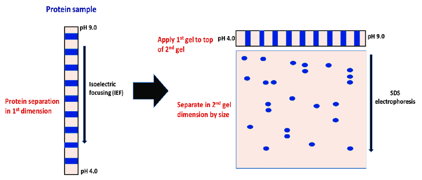
The extracted proteins are mixed with a loading dye and heated to denature them. They are then loaded into wells of a polyacrylamide gel and subjected to electrophoresis. SDS-PAGE separates proteins based on their molecular weight.
Transfer to Membrane
Once separated, proteins are transferred from the gel onto a membrane (usually nitrocellulose or PVDF) using electroblotting. The membrane captures the protein bands and provides a stable surface for further processing.
Blocking the Membrane
To prevent non-specific binding of probes, the membrane is blocked with a protein-rich solution (commonly BSA or non-fat milk in buffer). This ensures that the detection reagents will bind only to the target molecules.
Probing with Detection Molecules
This is the core of the Eastern blot. The membrane is incubated with specific detection reagents such as:
Lectins
Lectins are sugar-binding proteins used to detect glycosylated proteins.
Anti-Phosphorylation Antibodies
Used to detect phosphorylation, especially on tyrosine, serine, or threonine residues.
Lipid-Specific Probes
Lipid-binding dyes or antibodies detect lipidation like palmitoylation or myristoylation.
Enzymes and Chemical Tags
These bind to specific chemical groups on modified proteins.
Washing
The membrane is washed multiple times with buffer solutions to remove any unbound or loosely bound detection reagents, reducing background noise.
Detection
Depending on the type of probe used, various detection methods may be applied:
Colorimetric Detection
Enzyme-linked reactions produce a visible color.
Chemiluminescence

Enzyme reactions emit light that is captured on photographic film or digital imaging systems.
Fluorescence
Fluorophore-labeled probes emit light when excited with specific wavelengths.
Analysis
The resulting blot shows bands or spots corresponding to the presence of modified proteins. The size and intensity help identify which proteins are modified and to what extent.
Results and Interpretation
The final blot is analyzed by observing the appearance of bands or spots on the membrane. These bands indicate the presence of a specific modification on a protein of a particular size.
Key Points to Interpret
- Position of Bands: Corresponds to the molecular weight of the protein.
- Intensity of Bands: Indicates how much of the modified protein is present.
- Presence or Absence: Tells whether a specific modification exists in the sample.
Use of Controls
Controls are often included such as unmodified protein or a sample treated with an enzyme that removes the modification—to validate the detection’s specificity.
Applications of Eastern Blot
Eastern blotting is a powerful tool in specialized fields of research. Here are the key applications:
Detection of Glycosylation Patterns
Glycosylation is crucial for protein folding, stability, and signaling. Eastern blot helps detect and study these sugar modifications.
Study of Lipidated Proteins
Protein-lipid modifications impact membrane interactions. Eastern blot helps identify these changes.
Research on Phosphorylation
Eastern blot allows targeted detection of phosphorylated proteins using specific probes or chemical methods.
Quality Control in Biopharmaceuticals
Protein drugs require proper glycosylation. Eastern blotting ensures the accuracy of these modifications.
Identification of Disease Biomarkers
Altered protein modifications, especially glycosylation, are common in diseases like cancer. Eastern blotting can detect such biomarkers.
Vaccine Development
Pathogens often modify their surface proteins. Eastern blotting helps researchers study these modifications for better vaccine design.
Plant and Animal Research
Used to study stress responses and developmental processes in plants and animals by examining protein modifications.
Complementing Western Blotting
Eastern blotting can be used alongside Western blotting to give a more complete picture of protein identity and modifications.
Cancer Research
Changes in protein glycosylation and phosphorylation are common in cancer. Eastern blot helps researchers understand these patterns.
Limitations of Eastern Blot
While powerful, Eastern blotting has certain limitations:
Lack of Standardization
There are no universally accepted protocols for Eastern blotting, which can lead to inconsistencies.
Specialized Reagents Required
Probes for detecting modifications (e.g., lectins, antibodies) can be expensive or difficult to obtain.
Challenging Interpretation
Without proper controls and experience, it may be hard to interpret results accurately.
Limited Routine Use
Unlike Western blotting, Eastern blotting is not widely used in clinical laboratories due to complexity and specificity.
Conclusion
Eastern blot is a specialized technique that provides deep insights into the modifications of proteins after they are produced. While Western blot tells us what the protein is, Eastern blot reveals what has been added to it—such as sugars or lipids—that influence its function and behavior.
Though it’s not as commonly used as Western blot, Eastern blotting is crucial in fields such as cancer research, vaccine development, glycoproteomics , and drug manufacturing. As our understanding of post-translational modifications grows, Eastern blotting is likely to gain more attention and wider usage.
FREQUENTLY ASKED QUESTIONS
What is the main difference between Eastern and Western blot?
Western blot detects proteins using antibodies, while Eastern blot detects post-translational modifications like sugars or lipids using lectins or specific chemical probes.
Why is Eastern blot not as common as other blotting methods?
It is more specialized, lacks standard protocols, and often requires expensive reagents, limiting its routine use.
Can Eastern blot detect multiple types of modifications at once?
Usually, it focuses on one type of modification per blot. However, multiple probes can sometimes be used with proper planning.
Related Articles

