Introduction
Microbiology is a field that often deals with invisible life forms – bacteria, fungi, protozoa, and viruses. Among the various methods used to study these organisms, one fundamental technique is the direct microscopic count. This technique allows scientists and microbiologists to observe and count cells directly under a microscope. It is simple, quick, and very useful, especially in clinical, research, and food microbiology.
This document will walk you through every aspect of direct microscopic counts—its principle, procedure, types, applications, advantages, disadvantages, and much more, in a humanized and easy-to-understand format.
Table of Contents
What is Direct Microscopic Count?
Definition of Microscopic Counts
Direct Microscopic Count (DMC) is a microbiological method that involves the visual counting of cells using a microscope and a specialized slide, such as a counting chamber or hemocytometer. The method provides an immediate estimate of the number of microorganisms in a sample, whether it’s a culture, fluid, food item, or clinical specimen.
Why is microscopic Counts Important?
DMC is especially useful when:
- A rapid estimate of microbial numbers is needed.
- Observing morphology, arrangement, or motility of organisms.
- Counting viable and non-viable organisms in clinical or environmental samples.
Principle of Direct Microscopic Count
The basic principle of DMC relies on counting microorganisms in a known volume of a sample under a microscope using a grid-based slide. The number of organisms observed in a specific area is extrapolated to estimate the total number per milliliter (or per gram) of the sample.
Key Concepts of Microscopic Counts
- The microscope allows magnified visualization of microbes.
- A known area and depth under the coverslip allows calculation of volume.
- The number of cells per field is counted and used in calculations.
- Staining may be used to improve visibility and contrast of cells.
- Results are often expressed as cells/mL or cells/g.
Formula Used for Microscopic Counts
If you know the number of cells counted in a known volume, you can estimate the number of cells per mL using the following formula:
Number of cells/mL = (Average number of cells per square) × (Dilution factor) × (Conversion factor depending on chamber volume)
Instruments and Materials Required for Microscopic Counts
Microscope
- Typically a compound microscope with 400x–1000x magnification.
- Phase contrast or fluorescence microscopes may be used for specialized counts.
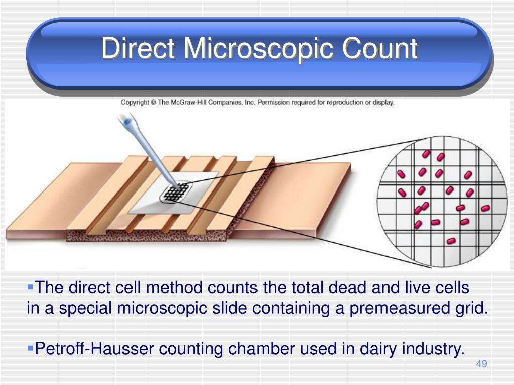
Counting Chamber (Hemocytometer)
- A hemocytometer is a precision-engineered glass slide with a grid etched into the center.
- It has known dimensions to calculate the exact volume under observation.
Coverslip
Special thick coverslips that are used with the hemocytometer to maintain the correct depth (usually 0.1 mm).
Pipettes
For accurate dilution and loading of the sample into the chamber.
Stains (Optional)
Methylene blue, crystal violet, Gram stain, or fluorescent dyes (like DAPI or acridine orange) can enhance visualization.
Procedure of Direct Microscopic Count
Step-by-Step Protocol of Microscopic Counts
Step 1: Sample Preparation
- Mix the sample well to ensure even distribution of organisms.
- If the sample is too concentrated, perform a serial dilution.
- For stained counts, mix the sample with the stain and wait for staining to complete.
Step 2: Charging the Hemocytometer
- Place the coverslip carefully over the grid.
- Using a pipette, load the sample into the V-shaped groove at the edge of the chamber.
- Capillary action will draw the fluid into the chamber evenly.
Step 3: Microscopic Observation
- Place the slide under the microscope.
- Adjust focus to visualize the grid lines clearly.
- Count the number of cells in specific squares (usually 5 large squares for averages).
Step 4: Calculating the Count
- Use the formula to calculate total cells per mL.
- Take averages if multiple squares are counted.
- Multiply by dilution factor if dilution was performed.
Types of Direct Microscopic Count Methods
1. Hemocytometer Method
- Common in research, blood cell counting, and yeast cell analysis.
2. Breed Smear Technique
- Used primarily in milk quality testing.
- A fixed volume of milk is spread on a slide, stained, and examined.
3. Petroff-Hausser Counting Chamber
- Specifically designed for bacterial counts.
- Has finer grid divisions than hemocytometers.
4. Epifluorescence Microscopy
- Utilizes fluorescent stains like DAPI.
- Used for marine, aquatic, and environmental samples.
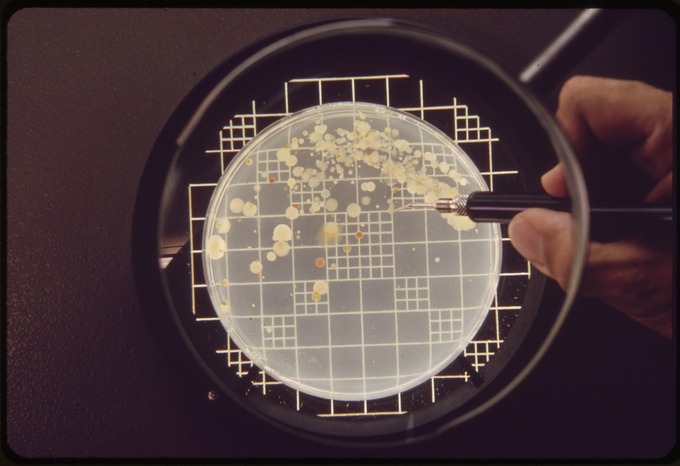
5. Membrane Filter Method
- A known volume of liquid is filtered, and the filter is placed on a slide.
- Cells on the membrane are stained and counted under a microscope.
Advantages of Direct Microscopic Count
Speed and Simplicity
Results are obtained quickly without the need for incubation.
Non-selectivity
All cells are counted, including dead, viable, and non-culturable ones.
Morphological Analysis
Cell size, shape, and arrangement can be observed simultaneously.
Useful for Non-culturable Organisms
Especially important in aquatic and clinical microbiology.
Limitations of Direct Microscopic Count
Can’t Differentiate Viable vs Non-viable Cells
Unless special staining is used (e.g., viability stains like trypan blue).
Human Error
Manual counting may lead to inconsistency and eye strain.
Not Suitable for Very Dilute Samples
If the sample has very few organisms, counts may be inaccurate.
Overlapping Cells
May cause miscounts due to clumping or poor preparation.
Applications of Direct Microscopic Counts
1. Clinical Microbiology
- Sputum, urine, CSF, and blood smears for immediate pathogen load estimation.
- Helps in disease diagnosis and treatment planning.
2. Food and Dairy Industry
- Checking microbial load in milk, meat, and processed foods.
- Part of quality control procedures.
3. Environmental Microbiology
- Counting bacteria in soil, water, and wastewater samples.
- Useful for pollution monitoring and ecological studies.
4. Pharmaceutical Industry
- Estimating microbial contamination in raw materials and finished products.
- Ensuring sterility and GMP compliance.
5. Biotechnology and Fermentation
- Monitoring yeast or bacterial populations in bioreactors.
- Helps in process optimization and yield prediction.
Comparisons with Other Counting Methods
vs Plate Count Method
- Plate counts only include viable organisms; DMC includes all.
- Plate count is more time-consuming but more specific.
vs Turbidity Method
- Turbidity measures optical density and needs calibration.
- DMC gives a visual and immediate count.
vs Flow Cytometry
- Flow cytometry is high-throughput and automated.
- DMC is manual, cheaper, and good for smaller scale work.
Accuracy and Reliability
How to Improve Accuracy?
- Count multiple fields and take an average.
- Avoid air bubbles and overloading the chamber.
- Use phase contrast microscopy for better contrast.
- Proper training and consistent technique help reduce errors.
Staining Techniques Used in DMC
1. Methylene Blue
Differentiates between live (unstained) and dead (blue) cells.
2. Gram Staining
Identifies Gram-positive vs Gram-negative bacteria.
3. Acridine Orange
Fluorescent stain for DNA and RNA, useful for environmental samples.
4. DAPI (4′,6-diamidino-2-phenylindole)
Binds to DNA, used in fluorescence microscopy.
5. Trypan Blue
Viability stain used for eukaryotic cells in culture.
Precautions and Tips
- Ensure uniform distribution of cells in the chamber.
- Use clean slides and coverslips to avoid background artifacts.
- Avoid drying out of the chamber during observation.
- Use counting aids or clickers to reduce eye strain and error.
Recent Advances and Innovations
Digital Microscopy
Automated systems that use image analysis software to count cells.
AI-Based Counting Systems
Machine learning models can improve speed and consistency.
Fluorescent-Based Kits
Specialized kits for quick DMC in food, blood, or environmental samples.
Conclusion
The Direct Microscopic Count is a fundamental yet powerful technique in microbiology. Despite its simplicity, it offers deep insights into the presence, abundance, and morphology of microorganisms in a sample. It continues to play a vital role in diagnostics, food safety, research, and industry.
Although newer automated and molecular techniques are emerging, DMC remains irreplaceable for rapid assessments, resource-limited settings, and when visual confirmation is needed.
FREQUENTLY ASKED QUESTIONS
Is Direct Microscopic Count accurate?
It is reasonably accurate when done carefully, but it can be affected by clumping, operator bias, or dilution errors.
Can DMC distinguish between live and dead cells?
Not directly. However, viability stains like trypan blue or methylene blue can help.
What is the most commonly used slide for DMC?
A hemocytometer or Petroff-Hausser counting chamber is widely used.
Related Articles