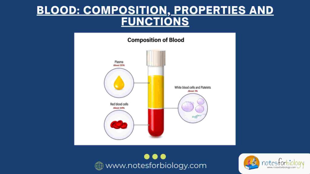
Cartilage is a specialized, semi-rigid connective tissue found throughout the human body. It plays vital roles in providing support, cushioning, and enabling smooth movements at joints. Unlike bone, cartilage is avascular, meaning it lacks a direct blood supply and depends on diffusion for nutrient exchange. Its unique composition and cellular arrangement make it essential in both embryonic development and adult tissue function.
Among the cellular components of cartilage, cartilage cells, or chondrocytes, are of particular importance. These cells are responsible for maintaining the cartilaginous matrix, participating in repair processes, and contributing to the overall physiology of connective tissues. Understanding the types, structure, examples, and functions of cartilage cells provides valuable insights into tissue biology and orthopedic health.
Summary of Cartilage Cells
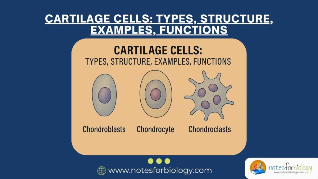
- Cartilage cells include chondrogenic cells, chondroblasts, and chondrocytes, each playing a role in cartilage formation, maintenance, and repair.
- Chondroblasts actively secrete the extracellular matrix, while mature chondrocytes maintain and regulate matrix turnover within lacunae.
- These cells support joint movement, absorb mechanical stress, and contribute to skeletal growth and repair, especially during embryonic development and at growth plates.
Table of Contents
Structure of Cartilage Cells
Before discussing the types, it is crucial to understand the general structure of cartilage cells. These cells possess distinct features that enable them to survive and function within the dense, resilient cartilage matrix.
Cartilage cells originate from precursor mesenchymal cells that differentiate into chondroblasts and later mature into chondrocytes. When active, these cells are typically rounded or oval in shape and are housed within small spaces called lacunae within the extracellular matrix. The matrix surrounding these cells is rich in collagen fibers and proteoglycans, giving cartilage its elastic and compressive strength. Each cartilage cell has a prominent nucleus and abundant cytoplasm containing organelles such as rough endoplasmic reticulum and Golgi apparatus, reflecting their active role in synthesizing cartilage matrix components. In mature cartilage, cells often appear in clusters known as isogenous groups, arising from the mitotic division of a single chondrocyte.
Types of Cartilage Cells
Cartilage tissue comprises several types of cartilage cells, classified based on their stage of differentiation and functional roles. Each type plays a critical part in cartilage development, maintenance, and repair. Broadly, there are three main types of cartilage cells found in the body.
Chondrogenic Cells
Chondrogenic cells are the cartilage-forming progenitor cells located in the inner layer of the perichondrium — a dense, fibrous membrane surrounding most cartilage. These cells are spindle-shaped and undifferentiated, possessing the potential to divide and differentiate into chondroblasts.
Their cytoplasm contains moderate amounts of organelles since their primary role is to act as a reserve population of precursor cells. Under appropriate physiological conditions, such as during cartilage growth or repair, these cells become active, giving rise to new chondroblasts and ensuring the continuity of cartilage tissue.
Chondroblasts
Chondroblasts represent the actively secreting form of cartilage cells. Derived from chondrogenic cells, they are located near the periphery of the cartilage tissue, particularly beneath the perichondrium. These cells are characterized by their rounded or ovoid shape and their abundance of rough endoplasmic reticulum, indicating their role in active protein synthesis.
Chondroblasts produce and secrete the main components of the extracellular matrix, including collagen fibers and proteoglycans. This matrix gradually surrounds the chondroblasts, isolating them into lacunae, at which point they mature into chondrocytes. Chondroblasts play a crucial role during the initial formation and growth of cartilage, as well as during tissue repair.
Chondrocytes
Chondrocytes are the mature, resident cells within cartilage tissue. Once enclosed in their lacunae, chondroblasts become chondrocytes, maintaining and modulating the cartilaginous matrix. These cells are responsible for balancing the synthesis and degradation of the matrix components to preserve cartilage integrity.
They are typically found singly or in small clusters called isogenous groups, indicating cell division after maturation. The cytoplasm of chondrocytes contains a well-developed rough endoplasmic reticulum and Golgi apparatus, essential for their ongoing secretory activity. Chondrocytes also respond to mechanical and biochemical stimuli, adapting the matrix composition to meet functional demands.
Examples of Cartilage Cells in Different Cartilage Types
Cartilage exists in the body in three major forms: hyaline cartilage, elastic cartilage, and fibrocartilage. The type of cartilage cells present and their organization vary depending on the cartilage type and its location.
In hyaline cartilage, the most common form found in the respiratory tract, articular surfaces, and embryonic skeleton, chondrocytes are present in round or oval lacunae within a homogenous matrix. These chondrocytes produce type II collagen and ground substances, ensuring resilience and support.
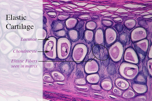
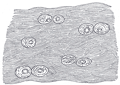
In elastic cartilage, present in the external ear, epiglottis, and Eustachian tube, chondrocytes are similarly located within lacunae but are surrounded by an extracellular matrix rich in elastic fibers. This composition imparts elasticity to the cartilage, allowing it to retain its shape after deformation.
Fibrocartilage, found in intervertebral discs and pubic symphysis, contains fewer chondrocytes embedded within a dense network of type I collagen fibers. The cells are often arranged in rows between collagen bundles, providing tensile strength and resistance to pressure.
Functions of Cartilage Cells
Cartilage cells play multiple essential roles in maintaining the structure and function of cartilaginous tissue. Their specific functions vary depending on their type and location within the body, but collectively, they contribute to the growth, maintenance, and repair of cartilage.
Matrix Production and Maintenance
A primary function of chondroblasts and chondrocytes is the production and maintenance of the extracellular matrix. This includes synthesizing collagen fibers and proteoglycans, which provide cartilage with its unique mechanical properties like elasticity, compressibility, and resistance to wear.
Chondroblasts actively secrete matrix components during cartilage formation and repair, while mature chondrocytes regulate matrix turnover by balancing synthesis and degradation to maintain tissue homeostasis.
Support and Shock Absorption
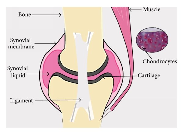
Chondrocytes indirectly contribute to support and shock absorption functions by maintaining the matrix’s structural integrity. The resilience of cartilage in absorbing mechanical stress in weight-bearing joints like the knees, hips, and vertebral column depends on the continuous activity of these cells.
By regulating the extracellular matrix composition, cartilage cells ensure that joints function smoothly and withstand repeated compressive and tensile forces without damage.
Growth and Development of the Skeleton
During embryonic development, cartilage cells play a crucial role in the formation of the initial cartilaginous model of the skeleton. This template later undergoes ossification in a process known as endochondral ossification, where cartilage is progressively replaced by bone.
Even in adults, cartilage cells are essential for bone growth at epiphyseal plates during childhood and adolescence, ensuring proper skeletal development and longitudinal bone growth.
Repair and Regeneration
Although cartilage has limited regenerative capacity due to its avascular nature, chondrocytes and chondrogenic cells participate in minor repair processes. In superficial injuries, surviving cartilage cells can proliferate and restore damaged matrix. However, larger defects are often replaced by fibrous tissue due to the restricted regenerative potential of cartilage.
Cartilage vs Bone
The comparative analysis between cartilage and bone highlights their distinct structural features, growth mechanisms, and functional roles within the body.
Composition and Structure
Cartilage is composed of chondrocytes within a gel-like matrix containing type II collagen and proteoglycans, while bone consists of osteocytes within a rigid, calcified matrix rich in type I collagen and hydroxyapatite crystals. Cartilage lacks blood vessels, nerves, and lymphatics, whereas bone is vascular and innervated.
Growth Mechanism
Cartilage grows by interstitial growth (from within by cell division) and appositional growth (from the outer layer). Bone, however, grows mainly by appositional growth through the addition of new layers by osteoblasts on existing surfaces.
Strength and Flexibility
Cartilage is flexible and compressible, making it suitable for cushioning and support in areas requiring movement. Bone is rigid and strong, providing mechanical protection and structural support.
Repair Capacity
Due to its avascular nature, cartilage has a poor regenerative capacity, while bone can heal effectively through a well-orchestrated repair process involving blood supply and osteoprogenitor cells.
Examples
- Cartilage: External ear, intervertebral discs, articular surfaces.
- Bone: Skull, femur, vertebrae, ribs.
Clinical Significance
Understanding the clinical relevance of cartilage cells and their associated tissues is essential for diagnosing, managing, and treating a variety of orthopedic and connective tissue diseases. Moreover, advances in regenerative medicine and tissue engineering continue to highlight the therapeutic potential of cartilage cells in modern healthcare.
Cartilage Disorders
Conditions like osteoarthritis involve the gradual degeneration of articular cartilage, leading to joint pain, stiffness, and reduced mobility. Chondrocyte dysfunction and matrix degradation are central to disease progression.
Congenital Anomalies
Developmental defects like achondroplasia result from impaired cartilage growth, causing dwarfism and skeletal abnormalities due to disrupted endochondral ossification.
Cartilage Injury and Repair
Cartilage injuries, particularly in athletes and aging individuals, heal poorly due to limited blood supply. Techniques like microfracture surgery, autologous chondrocyte implantation (ACI), and tissue engineering are employed to regenerate damaged cartilage.
Tissue Engineering Applications
Recent advances in cartilage tissue engineering involve culturing chondrocytes in biocompatible scaffolds to repair damaged cartilage in joints, the trachea, and other anatomical sites. Such applications hold promise for regenerative medicine and improving the quality of life in patients with cartilage defects.
Conclusion
In summary, cartilage cells are specialized, vital components of connective tissue that play indispensable roles in maintaining cartilage structure and function throughout life. Comprising chondrogenic cells, chondroblasts, and chondrocytes, these cells engage in matrix production, support, skeletal development, and repair.
Their varied morphology, location, and physiological roles contribute to the functional diversity of cartilage tissue types, including hyaline, elastic, and fibrocartilage. An in-depth understanding of cartilage cells not only enhances our knowledge of musculoskeletal biology but also informs clinical strategies in orthopedics, rheumatology, and tissue engineering for treating cartilage-related disorders.
Frequently Asked Questions (FAQ)
What are the main types of cartilage cells?
The main types of cartilage cells are chondrogenic cells, chondroblasts, and chondrocytes. Chondrogenic cells are stem-like precursors, chondroblasts actively produce the matrix, and chondrocytes are mature cells that maintain cartilage tissue.
What is the function of chondrocytes in cartilage?
Chondrocytes maintain and regulate the cartilage matrix, ensuring its strength, flexibility, and ability to absorb mechanical stress in joints and other structures.
Why does cartilage heal slowly?
Cartilage heals slowly because it is avascular (lacks a direct blood supply), so nutrients and repair cells reach cartilage through diffusion, limiting its regenerative capacity.