Centrioles and basal bodies are structurally related organelles found in most eukaryotic cells. Despite being small and often overlooked, they play crucial roles in cell division, motility, and organization of microtubules. Understanding their structure and functions offers deep insights into cellular processes and their implications in health and disease. This comprehensive overview explores the morphology, biogenesis, roles, and significance of centrioles and basal bodies in the cellular context.
1. Introduction
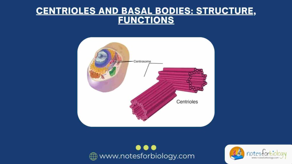
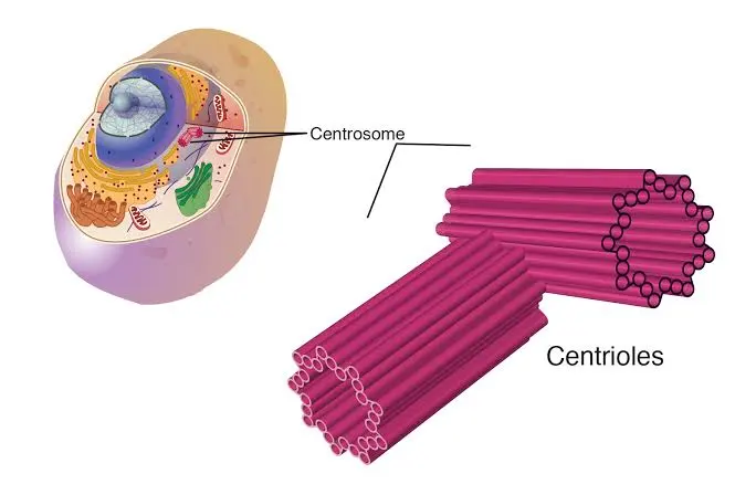
Centrioles and basal bodies are cylindrical structures composed primarily of microtubules. They are part of the microtubule organizing centers (MTOCs) and serve as core components in processes like mitosis, ciliogenesis, and maintenance of cellular architecture. Centrioles are typically found in pairs within the centrosome of animal cells, while basal bodies are found at the base of cilia and flagella. Though they differ in location and specific function, their structural similarities and interconnected roles suggest a common evolutionary origin.
Summary of Centrioles
- Centrioles are cylindrical structures made of nine triplet microtubules, located in the centrosome. They organize microtubules and support cell architecture.
- They direct the formation of the mitotic spindle during cell division, ensuring accurate chromosome segregation and genomic stability.
- Centrioles convert into basal bodies to form cilia and flagella, essential for cell motility and sensory functions. Defects are linked to diseases like cancer and ciliopathies.
Table of Contents
2. Structural Characteristics
2.1 Centrioles
Centrioles are cylindrical structures approximately 250 nm in diameter and 500 nm in length. Each centriole consists of nine triplet microtubules arranged in a cylindrical fashion. These triplets are labeled A, B, and C: A is a complete microtubule, while B and C share portions of their wall with A and B, respectively. This specific arrangement is termed the “9+0” configuration because there are no central microtubules.
Each centriole pair typically resides at right angles to each other in the centrosome, surrounded by pericentriolar material (PCM) that nucleates and anchors microtubules. The distal end of the centriole is associated with the formation of cilia or flagella, while the proximal end is where duplication begins during the cell cycle.
2.2 Basal Bodies
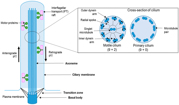
Basal bodies share the same “9+0” triplet microtubule arrangement as centrioles. In fact, basal bodies are often derived directly from centrioles. When a cell prepares to grow a cilium or flagellum, one of its centrioles migrates to the plasma membrane and becomes a basal body. The basal body anchors the axoneme, the central shaft of cilia and flagella, which extends from it and exhibits a “9+2” arrangement in motile forms.
Basal bodies are also associated with accessory structures such as rootlets and transition fibers, which help in anchoring and stabilizing the cilium or flagellum.
3. Biogenesis and Duplication
3.1 Centriole Duplication
Centrioles duplicate once per cell cycle to ensure proper centrosome inheritance. This duplication begins in the S phase when a new daughter centriole forms orthogonally to each mother centriole. This process is tightly regulated by cell cycle proteins like PLK4, SAS-6, STIL, and CEP135. Overexpression or mutation in these proteins can lead to abnormal centriole numbers, contributing to diseases like cancer.
The newly formed daughter centriole remains tightly associated with the mother centriole until the next cell cycle, during which they disengage and can form new pairs.
3.2 Basal Body Formation
Basal bodies can form through two major pathways:
- Centriole-dependent (template-based) pathway: In most vertebrate cells, basal bodies arise from pre-existing centrioles that migrate to the cell membrane and differentiate into basal bodies.
- De novo pathway: Seen in multiciliated cells, where hundreds of basal bodies form simultaneously in the absence of pre-existing centrioles. This requires specific proteins and scaffolding structures known as deuterosomes.
4. Functions
4.1 Role of Centrioles in Cell Division
Centrioles are essential for organizing the mitotic spindle during cell division. The centrosome, composed of two centrioles and the surrounding PCM, serves as the primary microtubule-organizing center (MTOC). During mitosis, centrosomes migrate to opposite poles of the cell and assist in forming the bipolar spindle apparatus.
Abnormalities in centriole duplication or function can lead to defective spindle formation, aneuploidy, and chromosomal instability, which are common features in many cancers.
4.2 Role of Basal Bodies in Cilia and Flagella Formation
Basal bodies nucleate the formation of cilia and flagella, acting as templates from which the axoneme grows. They anchor the cilia to the cell body and organize the internal structure necessary for ciliary movement. The basal body transitions into the axoneme by extending the A and B tubules of the triplet microtubules into doublets, forming the “9+2” structure typical of motile cilia.
Cilia and flagella serve various functions, including locomotion, fluid movement across epithelial surfaces, and signal transduction in sensory organs.
4.3 Non-Motile (Primary) Cilia
Non-motile, or primary cilia, typically have a “9+0” microtubule arrangement and lack the central pair found in motile cilia. These cilia are involved in sensing environmental signals and are important in pathways like Hedgehog, Wnt, and PDGF signaling.
Defective basal body function in primary cilia can result in developmental disorders known as ciliopathies, such as polycystic kidney disease, Bardet-Biedl syndrome, and Joubert syndrome.
5. Associated Proteins and Molecular Interactions
Centrioles and basal bodies rely on numerous associated proteins for their function and maintenance. Some key proteins include:
- PLK4 (Polo-like kinase 4): Master regulator of centriole duplication.
- SAS-6: Critical for cartwheel formation, establishing the ninefold symmetry of centrioles.
- CEP192, CEP135, and STIL: Involved in recruitment and stabilization of centriolar components.
- Gamma-tubulin: Part of the pericentriolar material, essential for microtubule nucleation.
- OFD1 and BBS proteins: Involved in basal body function and ciliogenesis.
These proteins interact in highly coordinated pathways to regulate centriole duplication, basal body formation, and cilia function.
6. Evolutionary Perspective
Centrioles and basal bodies are considered to have a common evolutionary origin, given their structural similarities and overlapping roles. They are conserved across a wide range of eukaryotic species, although some lineages like higher plants and certain fungi have lost centrioles, relying instead on alternative MTOCs.
The evolutionary conservation of centrioles and basal bodies highlights their fundamental importance in cellular organization and function. The transition from centrioles to basal bodies exemplifies evolutionary adaptation, allowing for the specialization of structures like cilia and flagella
7. Clinical Relevance and Diseases
Defects in centrioles and basal bodies are implicated in numerous diseases:
7.1 Cancer
Aberrant centriole duplication can result in supernumerary centrosomes, leading to multipolar spindles and chromosomal instability. This is a hallmark of many cancer cells. Mutations in centrosome-associated proteins like PLK4 and CEP192 are frequently observed in tumor samples.
7.2 Ciliopathies
As basal bodies are central to ciliogenesis, defects in their function lead to a group of disorders known as ciliopathies. These include:
- Polycystic Kidney Disease (PKD)
- Bardet-Biedl Syndrome (BBS)
- Meckel-Gruber Syndrome (MKS)
- Joubert Syndrome
These conditions often present with symptoms involving multiple organ systems, including the kidneys, liver, brain, and eyes.
7.3 Microcephaly and Developmental Disorders
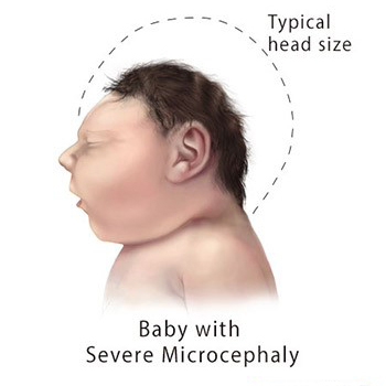
Mutations in centrosomal proteins can affect brain development, leading to microcephaly. The proper function of centrioles is crucial for neural progenitor cell division and brain size regulation.
8. Research and Technological Applications
Recent advances in microscopy and molecular biology have significantly enhanced our understanding of centriole and basal body biology. Techniques like super-resolution microscopy, cryo-electron tomography, and proteomics have allowed detailed visualization and identification of the protein composition of these structures.
Understanding centriole and basal body biology also has potential applications in regenerative medicine and synthetic biology. For example, manipulation of basal body function could aid in designing biomimetic systems or treating cilia-related disorders.
9. Conclusion
Centrioles and basal bodies, though small and once considered mere cellular appendages, are now recognized as vital components of cell structure and function. Their precise architecture and dynamic regulation underscore their importance in processes ranging from cell division to sensory reception. The implications of their dysfunction in disease contexts further highlight the necessity of continued research in this area. As science advances, the intricate biology of centrioles and basal bodies will undoubtedly remain a focal point for understanding both fundamental cellular mechanisms and complex pathological conditions.
Frequently Asked Questions (FAQs)
What are centrioles?
Centrioles are tiny cylindrical structures made of nine triplets of microtubules. They’re found in animal cells within the centrosome and are key for organizing the cell’s internal skeleton and helping cells divide.
What do centrioles do?
They help organize the mitotic spindle, which separates chromosomes during cell division, and act as precursors to basal bodies that build cilia and flagella.
How do centrioles duplicate?
During the S phase of the cell cycle, each centriole creates one new “daughter” centriole at a right angle. Duplication is tightly controlled to avoid random extra copies.
Related Articles