The human body is host to a diverse array of microorganisms, which form the normal microbial flora. These microorganisms play crucial roles in maintaining health and preventing pathogen colonization. The skin and throat are two areas with distinct microbial populations. This guide provides detailed procedures for isolating and identifying the normal flora of these regions.
Table of Contents
Normal microbial flora
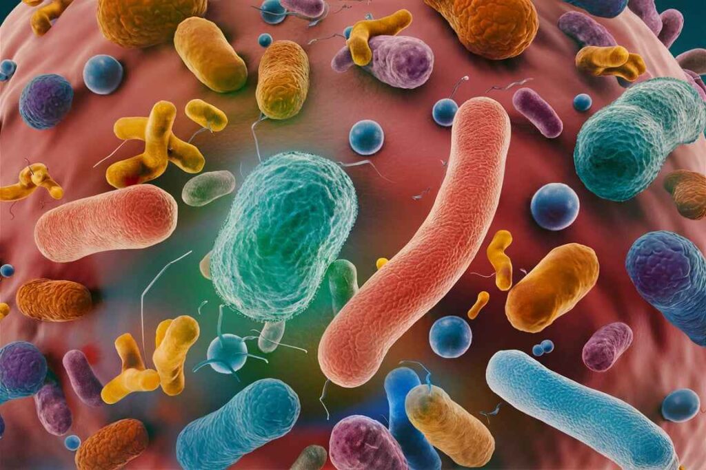
Normal microbial flora, also known as normal microbiota or microbiome, refers to the diverse community of microorganisms that reside on and within the bodies of healthy individuals. These microorganisms include bacteria, fungi, archaea, and viruses. Normal microbial flora typically inhabit various body sites, such as the skin, mouth, gastrointestinal tract, respiratory tract, and urogenital tract, without causing disease under normal conditions.
Skin Microbial Flora
Common Microbial Flora of the Skin
- Staphylococcus epidermidis: A major component of the skin flora, known for its coagulase-negative properties.
- Propionibacterium acnes: An anaerobic bacterium associated with sebaceous glands, playing a role in acne.
- Corynebacterium species: Non-pathogenic, diphtheroid bacteria.
- Micrococcus species: Common in exposed areas of the skin, usually harmless.
- Staphylococcus aureus: Sometimes part of the normal flora, more frequently found in nasal carriers.
Isolation Procedure
Sample Collection:
- Materials: Sterile cotton swabs, sterile saline solution, sterile test tubes.
- Procedure:
- Moisten a sterile swab with sterile saline.
- Swab the surface of the skin, such as the forearm or forehead, by rotating the swab over a 1-2 cm² area.
- Place the swab in a sterile test tube to transport it to the laboratory.
Culturing:
Agar Plates:
- Blood Agar: Supports growth of a wide range of bacteria and allows observation of hemolysis.
- Mannitol Salt Agar (MSA): Selective for staphylococci and differentiates S. aureus (ferments mannitol, producing yellow colonies).
- Nutrient Agar: General-purpose medium.
Inoculation:
- Streak the swab across the agar plates using a sterile technique.
Incubation:
- Incubate plates at 37°C for 24-48 hours.
Identification Procedure
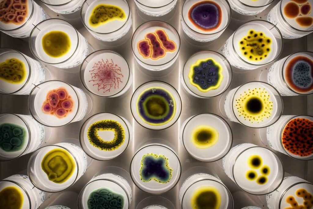
1. Colony Morphology:
- Examine colonies for size, shape, color, and hemolytic properties on blood agar.
- MSA will show yellow colonies if mannitol is fermented (indicative of S. aureus).
2. Gram Staining:
Procedure:
- Prepare a smear from a colony, heat-fix, and stain using Gram stain reagents.
- Observe under a microscope.
Interpretation:
- Gram-positive cocci in clusters suggest staphylococci.
- Gram-positive rods suggest corynebacteria.
3. Biochemical Tests:
Catalase Test:
- Drop hydrogen peroxide on a colony. Bubbling indicates a positive result.
- Staphylococci are catalase-positive; streptococci are catalase-negative.
Coagulase Test:
- Mix a colony with plasma. Clot formation indicates a positive result.
- S. aureus is coagulase-positive; other staphylococci are coagulase-negative.
Mannitol Fermentation:
- Observed on MSA. Yellow colonies indicate mannitol fermentation (S. aureus).
Throat Microbial Flora
Common Microbial Flora of the Throat
- Streptococcus viridans group: Dominant normal flora, alpha-hemolytic streptococci.
- Neisseria species: Non-pathogenic, part of normal flora.
- Haemophilus species: Present in the upper respiratory tract.
- Streptococcus pyogenes: Beta-hemolytic, sometimes present in healthy carriers.
Isolation Procedure
Sample Collection:
- Materials: Sterile cotton swabs, transport media.
- Procedure:
- Use a sterile swab to sample the posterior pharynx, avoiding contact with other mouth parts.
- Place the swab in transport media.
Culturing:
Agar Plates:
- Blood Agar: Supports a wide range of bacteria, showing hemolysis.
- Chocolate Agar: Enriched medium for fastidious organisms like Haemophilus and Neisseria.
- MacConkey Agar: Selective for Gram-negative bacteria.
Inoculation:
- Streak the swab across the agar plates using a sterile technique.
Incubation:
- Incubate plates at 37°C in a CO2-enriched environment for 24-48 hours.
Identification Procedure
Colony morphology:
On blood agar, observe colony features such as size, shape, color, and hemolysis (alpha, beta, or gamma).
Gram staining:
To stain a colony, prepare a smear, heat-fix it, and apply Gram stain reagents.
Examine under a microscope.
Interpretation:
Gram-positive cocci in chains indicate streptococci.
Gram-negative diplococci indicate Neisseria.
Biochemical tests:
- Catalase test distinguishes catalase-negative streptococci from catalase-positive staphylococci.
- To test optochin sensitivity, place a disk on a blood agar plate containing alpha-hemolytic bacteria.
- The zone of inhibition shows S. pneumoniae.
- To test bacitracin sensitivity, place a disk on a blood agar plate containing beta-hemolytic colonies.
- The zone of inhibition shows S. pyogenes.
- To do an oxidase test, apply the reagent to a colony. A hue change to dark purple implies a great outcome.
- Identifies the Neisseria species.
- The X and V Factor Test identifies Haemophilus species based on their development needs for hemin (X factor) and NAD (V factor).
Frequently Asked Question
Define Normal microbial flora.
Normal microbial flora, also known as normal microbiota or microbiome, refers to the diverse community of microorganisms that reside on and within the bodies of healthy individuals. These microorganisms include bacteria, fungi, archaea, and viruses.
What are the Common Microbial Flora of the Skin?
The Common Microbial Flora of the Skin are
1. Taphylococcus epidermidis
2. Propionibacterium acnes
3. Corynebacterium species
4. Micrococcus species
5. Staphylococcus aureus
Related Article
Mechanism of Plasmid replication: theta and rolling circle DNA replication