Nephrons are the microscopic structural and functional unit of the kidney responsible for the processes of filtration, reabsorption, secretion, and excretion. It is a long, coiled tubular structure, beginning in the renal cortex and extending into the renal medulla, which processes blood-derived filtrate through several specialized segments. Every function carried out by the kidney from the removal of nitrogenous wastes to the maintenance of electrolyte balance is made possible through the coordinated activity of millions of nephrons. Each human kidney contains approximately one to one and a half million nephrons, and together they ensure the purification and regulation of the body’s internal fluid environment.
Summary of Nephrons
- Nephrons are the functional units of the kidney that filter blood and form urine.
- There are two types: cortical and juxtamedullary nephrons, differing in location and role in urine concentration.
- Nephrons carry out urine formation through filtration, reabsorption, and secretion, helping regulate the body’s water, waste, and electrolyte balance.
Table of Contents
Location of Nephrons
Nephrons are situated within both the renal cortex and renal medulla of the kidney. The initial portions, including the renal corpuscle and proximal convoluted tubule, lie within the cortex. Parts of the nephron, such as the loop of Henle, extend into the medulla, while other components, like the distal convoluted tubule, return to the cortex. This arrangement allows for the efficient exchange of substances between the nephron and the surrounding capillary networks.
Structural Anatomy of Nephron
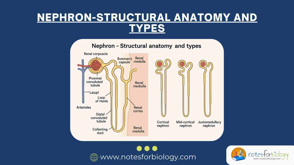
The nephron is composed of two principal components: the renal corpuscle and the renal tubule. Each of these components has distinct parts that contribute to the nephron’s function.
Renal Corpuscle
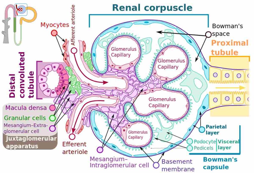
The renal corpuscle is the initial, spherical, blood-filtering structure of the nephron located in the renal cortex. It comprises two main parts: the glomerulus and the Bowman’s capsule.
Glomerulus
The glomerulus is a dense, tuft-like network of capillaries formed by the branching of an afferent arteriole. These capillaries are lined with fenestrated endothelial cells that allow the passage of water and small solutes while restricting the movement of larger molecules such as plasma proteins and blood cells. The high hydrostatic pressure within these capillaries forces fluid and dissolved substances out of the blood and into the surrounding Bowman’s capsule.
Bowman’s Capsule
Encasing the glomerulus is the Bowman’s capsule, a double-walled, cup-shaped epithelial structure. It consists of an outer parietal layer made of simple squamous epithelium and an inner visceral layer composed of specialized cells called podocytes. These podocytes have interdigitating foot-like extensions (pedicels) that create narrow filtration slits, further regulating the filtration process. The space between the two layers is known as Bowman’s space or capsular space, where the glomerular filtrate is initially collected.
Renal Tubule
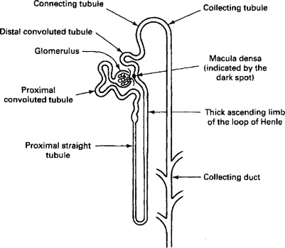
Following the renal corpuscle, the filtrate passes into the renal tubule, a long, convoluted structure that processes the filtrate into urine by reabsorbing valuable substances and secreting additional waste products.
Proximal Convoluted Tubule (PCT)
The proximal convoluted tubule is the first segment of the renal tubule, arising from Bowman’s capsule. It is located in the renal cortex and is characterized by a highly coiled structure. The PCT is lined with simple cuboidal epithelium featuring an abundance of microvilli, which form a brush border to increase the surface area for reabsorption. This segment is responsible for the reabsorption of a majority of the filtrate, including glucose, amino acids, ions, and water.
Loop of Henle
From the proximal convoluted tubule, the filtrate enters the Loop of Henle, a U-shaped segment that extends from the cortex into the medulla and back to the cortex. It consists of three parts: the descending limb, the thin ascending limb, and the thick ascending limb.
The descending limb is permeable to water but relatively impermeable to solutes, allowing water to be reabsorbed into the surrounding interstitial fluid.
The thin ascending limb has limited permeability to water but allows for the passive movement of sodium and chloride ions out of the tubular fluid.
The thick ascending limb is impermeable to water and actively transports sodium, potassium, and chloride ions from the tubular fluid into the surrounding medullary interstitium, contributing to the formation of an osmotic gradient necessary for urine concentration.
Distal Convoluted Tubule (DCT)
After the Loop of Henle, the filtrate enters the distal convoluted tubule, situated in the renal cortex. The DCT is shorter and less convoluted than the PCT and is lined with simple cuboidal epithelial cells with fewer microvilli. The DCT plays a crucial role in the selective reabsorption of sodium and calcium ions and the secretion of potassium, hydrogen, and ammonium ions. It is also influenced by hormones such as aldosterone and parathyroid hormone, which regulate electrolyte balance.
Collecting Duct
The final segment of the nephron system is the collecting duct, which receives urine from multiple distal convoluted tubules. The collecting ducts travel through the renal medulla, converging with other ducts to form larger papillary ducts that open into the minor calyces. The collecting duct system is lined with principal and intercalated cells and is involved in the final regulation of water, sodium, and acid-base balance under the influence of antidiuretic hormone (ADH) and aldosterone.
Juxtaglomerular Apparatus (JGA)
Associated with the nephron is a specialized structure known as the juxtaglomerular apparatus, located near the renal corpuscle where the distal convoluted tubule makes contact with the afferent arteriole. The JGA plays a vital role in the regulation of blood pressure and glomerular filtration rate.
The juxtaglomerular apparatus consists of:
- Juxtaglomerular cells, which are modified smooth muscle cells of the afferent arteriole that produce and secrete the enzyme renin in response to decreased blood pressure.
- Macula densa cells, which are specialized epithelial cells in the distal convoluted tubule that sense sodium chloride concentration in the filtrate.
- Extraglomerular mesangial cells, which provide structural support and mediate communication between the juxtaglomerular cells and macula densa.
Types of Nephrons
Based on their location, structure, and function, nephrons are classified into two distinct types: cortical nephrons and juxtamedullary nephrons. These types differ in their anatomical position and their role in urine concentration.
Cortical Nephrons
Cortical nephrons constitute approximately 85% of the total nephron population in the human kidney. They are primarily located in the outer cortex of the kidney, with relatively short loops of Henle that barely extend into the outer medulla. The main function of cortical nephrons is to carry out the majority of filtration, reabsorption, and secretion. Due to their short loops of Henle, they play a limited role in establishing the medullary osmotic gradient necessary for the concentration of urine.
Juxtamedullary Nephrons
Juxtamedullary nephrons account for about 15% of the total nephrons. They are located close to the corticomedullary junction and possess long loops of Henle that extend deep into the inner medulla, sometimes reaching the renal papilla. These nephrons are primarily responsible for the generation of a concentrated urine by creating and maintaining a steep osmotic gradient within the medullary interstitium. The long loops of Henle, along with their specialized capillary network called the vasa recta, enable effective countercurrent exchange mechanisms essential for water conservation and urine concentration.
Differences Between Cortical and Juxtamedullary Nephrons
Let’s organize the differences in a clear comparison.
| Feature | Cortical Nephrons | Juxtamedullary Nephrons |
|---|---|---|
| Number | 85% of total nephrons | 15% of total nephrons |
| Location of Renal Corpuscle | Outer cortex | Near corticomedullary junction |
| Length of Loop of Henle | Short | Long (extends deep into the medulla) |
| Presence of Vasa Recta | Poorly developed or absent | Well-developed and extensive |
| Role in Urine Concentration | Minimal | Major role through counter-current system |
| Function | Basic filtration, reabsorption, secretion | Concentration of urine, water conservation |
Nephron and Urine Formation
The nephron carries out urine formation in three primary stages: glomerular filtration, tubular reabsorption, and tubular secretion.
Glomerular filtration
It occurs in the renal corpuscle, where plasma is filtered under high pressure through the glomerular capillaries into Bowman’s capsule. This process forms an ultrafiltrate devoid of blood cells and large proteins but rich in water, glucose, ions, and nitrogenous wastes.
Tubular reabsorption
It is the selective recovery of essential substances from the filtrate back into the bloodstream. It occurs primarily in the proximal convoluted tubule, where about 65-70% of water, glucose, amino acids, and electrolytes are reclaimed. Additional reabsorption happens in the loop of Henle, distal convoluted tubule, and collecting duct.
Tubular secretion
It involves the active transport of additional waste products and ions from the blood into the tubular fluid. This process occurs in the proximal and distal convoluted tubules and collecting ducts, ensuring the elimination of substances such as hydrogen ions, potassium, creatinine, and certain drugs.
Through these stages, the nephron transforms blood plasma into urine while precisely adjusting the body’s water, electrolyte, and acid-base balance.
Nephron Pharmaceuticals
Nephron Pharmaceuticals refers to therapeutic agents specifically designed to act on various parts of the nephron to treat kidney-related and systemic conditions. These drugs target specific nephron segments to alter reabsorption and secretion processes, thereby controlling blood pressure, electrolyte levels, and fluid volume.
Diuretics are among the most well-known nephron-targeting drugs. Loop diuretics like furosemide act on the thick ascending limb of the Loop of Henle to inhibit sodium, potassium, and chloride reabsorption, resulting in increased urine output. Thiazide diuretics work on the distal convoluted tubule to reduce sodium reabsorption, while potassium-sparing diuretics affect the collecting duct, minimizing potassium loss.
Renin-Angiotensin-Aldosterone System (RAAS) inhibitors, including ACE inhibitors and angiotensin receptor blockers (ARBs), indirectly influence nephron function by reducing the effects of aldosterone, thereby decreasing sodium and water reabsorption.
Other drugs such as carbonic anhydrase inhibitors, antidiuretic hormone antagonists, and phosphate binders also target nephron structures to manage specific clinical conditions like glaucoma, heart failure, and chronic kidney disease.
Conclusion
The nephron is an intricately structured and highly specialized unit that enables the kidneys to perform their vital physiological functions. Comprising the renal corpuscle and renal tubule, the nephron executes complex processes of filtration, reabsorption, secretion, and excretion to maintain homeostasis. The presence of two distinct types of nephrons cortical and juxtamedullary — allows for the efficient regulation of blood composition and urine concentration according to the body’s requirements.
An understanding of the nephron’s structure and types provides essential insight into kidney physiology and the mechanisms underlying renal function and disorders. This intricate organization underscores the remarkable ability of the kidneys to maintain the internal environment in a balanced and regulated state.
Frequently Asked Questions (FAQ)
Why nephron is the functional unit of kidney?
The nephron is the functional unit of the kidney because it performs all the essential filtration and purification processes that remove waste and excess fluids from the blood, ultimately producing urine.
What is a nephron and where is it located?
A nephron is the microscopic structural and functional unit of the kidney responsible for filtering blood and forming urine. It is located in both the renal cortex and medulla, with different parts of the nephron extending through these kidney regions.
Are nephron cells.
No, nephrons are not individual cells, but rather the functional units of the kidney.