The embryonic development of the frog is one of the most widely studied processes in developmental biology. This is because frogs have external fertilization, and their transparent embryos develop outside the mother’s body, making the various developmental stages easy to observe. The process of development begins with fertilization and proceeds through cleavage, blastula formation, gastrulation, neurulation, organogenesis, tailbud formation, and finally, hatching into a free-swimming tadpole.
Summary of Key Processes & Structures
| Stage | Main Events and Structures |
|---|---|
| Fertilization | Sperm-egg fusion, gray crescent formation |
| Cleavage | Holoblastic unequal divisions, morula formation |
| Blastula | Formation of blastocoel |
| Gastrulation | Formation of three germ layers, archenteron |
| Neurulation | Formation of neural tube and notochord |
| Organogenesis | Differentiation into organs and tissues |
| Tailbud stage | Embryo elongation, heart starts beating |
| Hatching | Tadpole emerges, begins independent life |
Table of Contents
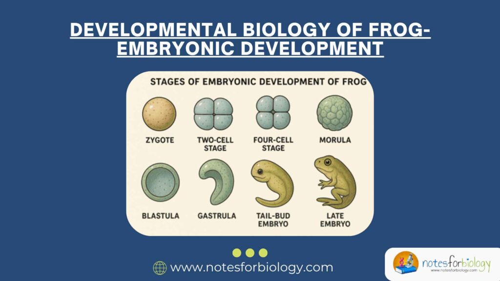
Stages of Embryonic Development of Frog
Fertilization
Fertilization in frogs is external and takes place in water. During mating, the male frog clasps the female in a position known as amplexus and releases sperm over the eggs as the female lays them in water. The fusion of a single sperm with an egg initiates fertilization. After the sperm penetrates the egg membrane, a fertilization membrane forms around the egg to prevent the entry of additional sperm, a phenomenon known as the block to polyspermy.
A significant feature of frog fertilization is the formation of the gray crescent, a light-colored, crescent-shaped region that appears on the surface of the egg opposite the point of sperm entry. This gray crescent marks the future dorsal side of the embryo and is crucial for establishing the body’s primary axes (anterior-posterior, dorsal-ventral, and left-right). The egg itself is polarized into an animal pole (upper, darker region with less yolk) and a vegetal pole (lower, lighter region rich in yolk).
Cleavage
Following fertilization, the zygote undergoes a series of rapid, mitotic cell divisions known as cleavage. In frogs, cleavage is holoblastic (complete) but unequal, due to the presence of a large amount of yolk in the vegetal pole, which slows down cell division there. The first cleavage is a vertical division passing through the animal and vegetal poles, dividing the egg into two equal blastomeres. The second cleavage is also vertical but occurs at a right angle to the first. The third cleavage is horizontal but unequal, producing smaller cells (micromeres) in the animal pole and larger cells (macromeres) in the vegetal pole.
As cleavage continues, a solid ball of cells called a morula is formed. The morula then reorganizes into a hollow, spherical structure called the blastula. The cavity within the blastula is known as the blastocoel, and it is eccentrically located towards the animal pole due to the uneven distribution of yolk.
Blastula Formation
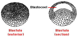
The blastula stage marks the end of cleavage and the beginning of major morphogenetic movements. It is characterized by the formation of a single-layered hollow ball of cells enclosing the blastocoel. At this stage, the cells at the animal pole continue to divide more rapidly than those at the vegetal pole. The blastocoel provides a space for cell movements during the next stage, gastrulation. The blastula is still surrounded by a fertilization membrane, which will eventually break down.
Gastrulation
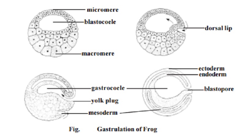
Gastrulation is one of the most critical phases in embryonic development as it leads to the formation of the three primary germ layers: ectoderm, mesoderm, and endoderm. It begins with the appearance of a depression on the surface of the embryo at the gray crescent region. This depression, known as the blastopore, marks the initiation of complex cellular movements.
Cells move inward through the blastopore by invagination (inward bending of a cell sheet) and involution (movement of cells over the edge of the blastopore into the embryo’s interior). The blastopore’s upper edge, called the dorsal lip of the blastopore, is especially important because it acts as the primary organizer of development.
As gastrulation proceeds, the blastocoel gradually shrinks and is replaced by a new cavity called the archenteron, the primitive gut. The archenteron’s opening to the exterior is the blastopore, which will later become the anus. At this stage, the embryo is known as a gastrula. The ectoderm covers the outside, the mesoderm lies in the middle, and the endoderm lines the archenteron.
Neurulation
After gastrulation, the process of neurulation begins. This stage is characterized by the formation of the nervous system, particularly the neural tube, which will develop into the brain and spinal cord. Neurulation starts when a region of the ectoderm above the notochord, called the neural plate, thickens and forms a flat sheet of cells. The edges of this plate rise to form the neural folds, and the depression between them is called the neural groove.
As development continues, the neural folds converge towards the midline and fuse, transforming the neural groove into the neural tube. This tube detaches from the overlying ectoderm and sinks into the body to eventually form the central nervous system. Cells at the tips of the neural folds, known as neural crest cells, migrate to different parts of the embryo and give rise to various structures such as peripheral nerves, pigment cells, and facial cartilage.
Organogenesis
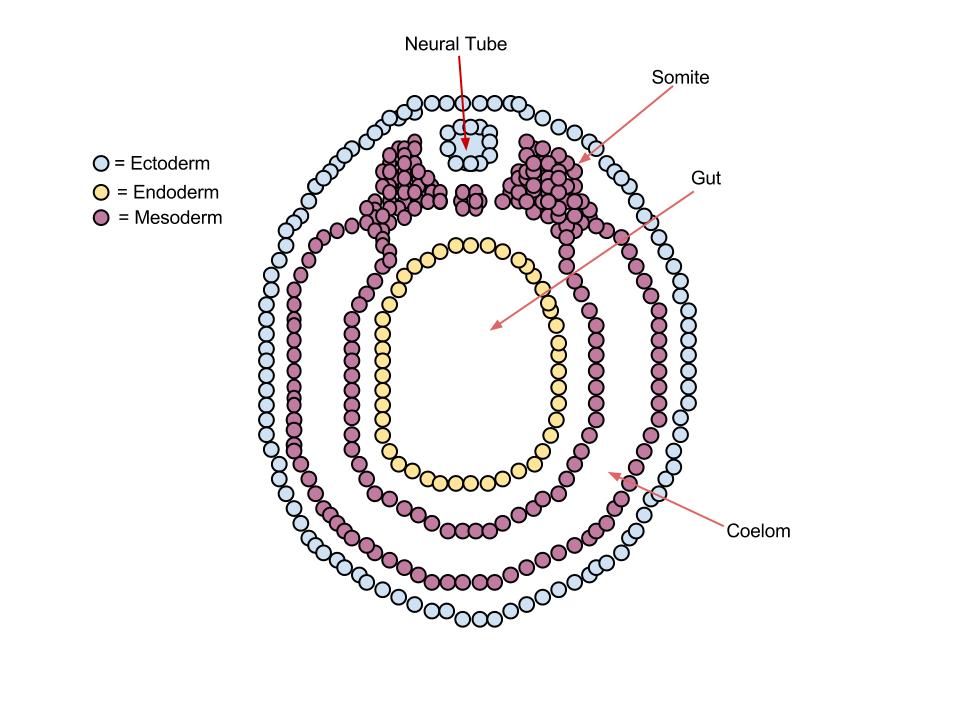
Once the basic body plan has been established through gastrulation and neurulation, the embryo enters the organogenesis stage. During this phase, the three germ layers differentiate into various tissues and organs.
- The ectoderm gives rise to the epidermis (outer skin) and the entire nervous system, including the brain, spinal cord, and nerves.
- The mesoderm forms the notochord, muscles, skeleton, circulatory system, kidneys, gonads, and connective tissues. The mesoderm also segments into structures called somites, which will later develop into vertebrae and associated muscles.
- The endoderm differentiates into the lining of the digestive tract, respiratory system, and organs such as the liver and pancreas.
This stage involves both the growth and functional differentiation of cells into specialized tissues.
Tailbud Stage
Following organogenesis, the embryo progresses to the tailbud stage, during which the overall body shape becomes more elongated, and structures like the tailbud and gills become visible. The head region enlarges, and the eyes begin to form as bulges on either side of the head. The heart starts to beat, and blood circulation begins, ensuring the transport of nutrients and oxygen to the rapidly developing tissues.
The neural tube continues to differentiate into distinct brain regions and the spinal cord. The tail, an essential feature for swimming in the aquatic environment, extends and takes shape.
Hatching
After the completion of the tailbud stage, the embryo is ready to hatch. The developing tadpole secretes enzymes that help it break through the surrounding egg membrane. Once free, the tadpole swims actively in the water, feeding on algae and continuing its growth and development. The tadpole possesses gills for respiration, a long tail for swimming, and a simple digestive system suited to a herbivorous diet. It will later undergo metamorphosis to develop into an adult frog, a process involving the resorption of the tail, development of limbs, and transition from gills to lungs for respiration.
Significance of Frog in Developmental Biology
Frogs, especially the African clawed frog (Xenopus laevis), have long been considered model organisms in developmental biology. Their unique biological characteristics and practical experimental advantages have made them valuable tools for understanding the fundamental processes of early vertebrate development. Here’s a detailed explanation of their significance:
Easy Availability and Large, Transparent Eggs
One of the major advantages of using frogs in developmental studies is the large size of their eggs, which are visible to the naked eye and easy to handle in laboratory conditions. The transparent nature of the eggs’ outer layers in some species allows scientists to observe embryonic development in real time, from fertilization to hatching. This makes it possible to track early cleavage patterns, gastrulation movements, and organ formation directly under a microscope without invasive procedures.
External Fertilization and Rapid Development
Frogs undergo external fertilization, which simplifies the collection of large numbers of synchronized embryos at specific stages. This is particularly useful for experiments requiring uniform developmental stages for observation or manipulation. Moreover, frogs exhibit rapid embryonic development, with major structures such as the neural tube and organ rudiments forming within just a few days, allowing for quick experimental turnarounds and developmental tracking.
Historical Contributions to Embryology Concepts
Frog embryos played a crucial role in the discovery and understanding of several fundamental principles in developmental biology:
- Organizer Concept: The famous Spemann and Mangold experiment (1924) used frog embryos to demonstrate the existence of the primary organizer region (the dorsal lip of the blastopore). When transplanted, this region could induce the formation of a second body axis in a host embryo, leading to the discovery of embryonic induction.
- Gray Crescent: Studies on frog eggs led to the identification of the gray crescent region, essential for axis formation and proper gastrulation.
These landmark findings laid the foundation for modern concepts in embryology, such as morphogen gradients, cell fate determination, and induction signaling pathways.
Genetic and Molecular Research Advantages
In recent decades, Xenopus species have also become useful in molecular and genetic studies.
- Xenopus tropicalis, with a diploid genome and shorter generation time compared to Xenopus laevis, is increasingly used for genetic manipulation, including CRISPR-Cas9 gene editing.
- Frog oocytes (immature egg cells) are large and capable of protein synthesis, making them an ideal system for injecting mRNA, DNA, and proteins to study gene function, protein interactions, and cell signaling mechanisms in a controlled environment.
Biomedical and Evolutionary Insights
Frog embryos have contributed valuable insights into vertebrate evolution and comparative embryology. Since amphibians occupy a phylogenetic position between fish and higher vertebrates like reptiles, birds, and mammals, studying their development helps bridge gaps in understanding vertebrate evolution. Moreover, Xenopus embryos have been used to model human diseases, birth defects, and developmental disorders by manipulating genes and signaling pathways analogous to those in humans.
In Vitro Experimental Versatility
Frog embryos are exceptionally suitable for in vitro (outside the body) experiments. Scientists can remove and transplant tissues, isolate specific embryonic regions, and observe the effects of drugs, hormones, or environmental changes on embryonic development. This versatility makes them an indispensable tool for developmental biology research.
Conclusion
The embryonic development of the frog is a remarkable and well-organized process that involves a sequence of carefully orchestrated stages, starting from fertilization and culminating in the formation of a free-swimming tadpole. Each stage — from cleavage, blastulation, gastrulation, neurulation, organogenesis, to the tailbud and hatching stages — plays a critical role in establishing the frog’s body plan and organ systems. The process highlights essential concepts of developmental biology, such as cell division, differentiation, and morphogenesis. Studying frog embryology provides invaluable insights into the basic mechanisms of vertebrate development and serves as a model for understanding human embryogenesis.
Frequently Asked Questions (FAQ)
What is the Correct sequence in embryonic development of frog?
The development sequence begins with fertilization, followed by cleavage and blastula formation.
Then gastrulation, neurulation, and organogenesis take place.
Finally, the embryo forms a tailbud and hatches into a tadpole.
How is a frog embryo formed?
A frog embryo forms when a sperm fertilizes an egg externally in water, initiating cell divisions that gradually shape the embryo.
What type of fertilization occurs in frogs?
Frogs have external fertilization, where eggs and sperm meet in water.