Chromosomes are essential components of living cells, responsible for carrying the genetic material that determines the traits and biological processes of an organism. These thread-like structures play a crucial role in heredity, cellular function, and the regulation of genetic information within the cell nucleus.
From the early days of cytogenetics to modern genome editing techniques, chromosomes have remained at the center of genetic research. This article explores their definition, structure, types, functions, abnormalities, and research applications, providing a detailed understanding of their significance in biology and medicine.
Summary of Chromosomes
- Chromosomes are thread-like structures made of DNA and proteins that store and transmit genetic information, ensuring the inheritance of traits and regulating essential cellular activities.
- They have a defined structure with parts like the centromere, telomere, and chromatids, and are classified based on number, function (autosomes and sex chromosomes), and centromere position.
- Chromosomal abnormalities in number or structure can cause genetic disorders and diseases, and modern techniques like karyotyping, FISH, and CRISPR have advanced our ability to analyze, modify, and understand chromosomes in health, medicine, and evolution.
Table of Contents
Importance of Chromosomes in Genetics and Cellular Biology
In the study of genetics, chromosomes serve as the carriers of genes the fundamental units of heredity. They ensure the accurate transmission of genetic traits from parents to offspring and regulate essential cellular processes such as growth, development, and metabolism.
They are equally important in medical genetics, where chromosomal abnormalities are linked to various diseases and developmental disorders. Their structure and function continue to be studied to understand genetic inheritance, cellular behavior, and evolutionary biology.
Overview of Their Discovery and Research Significance
The discovery of chromosomes marked a pivotal moment in biology. First observed under the microscope in dividing cells during the 19th century, chromosomes were soon linked to heredity and gene function. Early cytologists like Walther Flemming, Wilhelm Roux, and Theodor Boveri laid the foundation for chromosomal theory.
In the 20th and 21st centuries, advances in microscopy, molecular biology, and genomics have revolutionized our understanding of chromosomes, leading to breakthroughs in gene mapping, cancer research, and genome editing technologies like CRISPR.
What are Chromosomes?
A chromosome is a highly organized structure composed of DNA and proteins, found in the nuclei of eukaryotic cells and within the cytoplasm of prokaryotic organisms. These structures house the genetic instructions necessary for life, encoded within sequences of nucleotides.
Definition of Chromosomes
They are defined as linear or circular DNA molecules associated with specific proteins, forming a compact structure that stores and transmits genetic information. In eukaryotes, chromosomes reside in the nucleus, whereas prokaryotic chromosomes are typically circular and located in the cytoplasm.
Each species has a characteristic number of chromosomes, collectively referred to as its karyotype. In humans, for example, the diploid number is 46, comprising 22 pairs of autosomes and one pair of sex chromosomes.
Etymology and Historical Discovery
The term chromosome derives from the Greek words chroma (color) and soma (body), coined by German anatomist Heinrich Wilhelm Waldeyer in 1888. This reflects the property of chromosomes to take up certain stains, making them visible under a microscope during cell division.
Walther Flemming first observed dividing chromosomes in the late 1800s, while subsequent researchers established their role in inheritance. The landmark work of Boveri and Sutton in the early 20th century led to the chromosome theory of inheritance, integrating chromosomes into Mendel’s laws of heredity.
Structure of Chromosomes
Chromosomes possess a well-defined structure, essential for the efficient packaging, protection, and regulation of genetic material. Their composition ensures stability during cell division and accessibility during gene expression and DNA repair.
Basic Components: DNA and Proteins
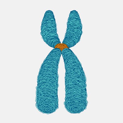
The primary component of a chromosome is DNA, the molecule that carries genetic instructions in a double helix structure. This DNA is tightly wound around histone proteins, forming complexes called nucleosomes.
The combination of DNA and histone proteins creates a chromatin structure that can condense into visible chromosomes during cell division. This organization prevents DNA damage and facilitates efficient gene regulation and replication.
Chromatin Organization: Euchromatin vs. Heterochromatin
Within the nucleus, chromatin exists in two main forms: euchromatin and heterochromatin. Euchromatin is loosely packed and transcriptionally active, allowing gene expression. In contrast, heterochromatin is densely packed, generally inactive, and often found at the chromosome’s periphery.
This functional division enables the cell to selectively access genetic information based on its developmental stage, cell type, and environmental conditions. Heterochromatin also plays a role in maintaining chromosome stability and protecting telomeres.
Parts of a Chromosome
Each chromosome is composed of several distinct regions and substructures that contribute to its function and stability.
Centromere
The centromere is a constricted region of the chromosome that serves as the attachment point for spindle fibers during cell division. It ensures the equal distribution of chromosomes to daughter cells by maintaining sister chromatid cohesion.
Centromeres are classified based on their position within the chromosome and can exhibit variations like metacentric, acrocentric, or telocentric forms. Their DNA sequence and associated proteins form a specialized chromatin structure known as kinetochore.
Telomere
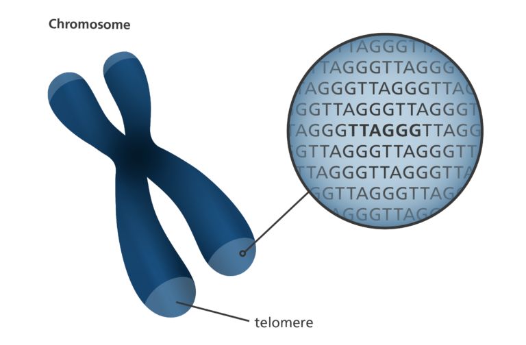
Telomeres are protective structures found at the ends of linear chromosomes, composed of repetitive non-coding DNA sequences (e.g., TTAGGG in humans). They prevent chromosome degradation, fusion with neighboring chromosomes, and the loss of genetic information during DNA replication.
Telomere length gradually shortens with each cell division, contributing to cellular aging and limiting the number of divisions a cell can undergo a phenomenon known as the Hayflick limit.
Chromatid
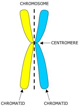
A chromatid is one of two identical halves of a duplicated chromosome, joined together at the centromere. During mitosis and meiosis, chromatids are separated and distributed into daughter cells, ensuring the faithful transmission of genetic material.
When joined, they are referred to as sister chromatids, while individual chromatids become independent chromosomes after separation.
Chromonema
The chromonema refers to the coiled thread-like structure formed by condensed chromatin during early stages of cell division. It represents a higher-order organization of nucleosome fibers and can be observed under a microscope as thin threads within the chromosome.
The chromonema undergoes further condensation to form the characteristic metaphase chromosome structure during mitosis and meiosis.
Matrix and Nucleosome Structure
The chromosomal matrix provides structural support and serves as a scaffold for chromatin fibers and regulatory proteins. It maintains the shape and organization of the chromosome, particularly during metaphase.
Chromatin is organized into repeating structural units called nucleosomes, each consisting of DNA wrapped around a histone octamer. This “beads-on-a-string” structure compacts the DNA by nearly sevenfold, enabling efficient packaging within the nucleus.
Types of Chromosomes
Chromosomes can be classified based on their roles in inheritance, structural similarities, and the position of their centromere. Understanding these classifications helps explain genetic behavior during cell division and the inheritance of traits.
Different types of chromosomes are essential for determining sex, controlling body functions, and maintaining the integrity of the genetic code during reproduction and development.
Autosomes vs. Sex Chromosomes
In most organisms, chromosomes are divided into two broad categories: autosomes and sex chromosomes. Autosomes are chromosomes that carry genes responsible for the general characteristics of an organism, excluding those that determine sex.
In humans, there are 22 pairs of autosomes and one pair of sex chromosomes, making a total of 46 chromosomes. The sex chromosomes determine an individual’s sex: typically XX in females and XY in males. These also carry genes unrelated to sex, contributing to overall health and development.
Homologous vs. Non-Homologous Chromosomes
Chromosomes also differ based on their genetic similarity. Homologous chromosomes are a pair of chromosomes, one inherited from each parent, that are similar in shape, size, and gene content. They pair during meiosis to ensure accurate segregation of genetic material.
Non-homologous chromosomes, on the other hand, are those that differ in size, structure, and gene content. These chromosomes do not pair with each other during meiosis and have distinct functions within the cell.
Based on Centromere Position
The physical appearance and classification of chromosomes often depend on the location of the centromere, which divides the chromosome into two arms.
Metacentric
A metacentric chromosome has its centromere positioned in the middle, resulting in two arms of equal length. These chromosomes form a characteristic V-shape during cell division and are commonly seen in human karyotypes.
Submetacentric
In submetacentric chromosomes, the centromere is slightly off-center, producing one short arm and one long arm. These chromosomes take on an L-shaped appearance during metaphase.
Acrocentric
An acrocentric chromosome has its centromere near one end, creating one very short arm and one very long arm. In humans, chromosomes 13, 14, 15, 21, and 22 are acrocentric and are known for containing satellite bodies or nucleolar organizer regions (NORs).
Telocentric
A telocentric chromosome has its centromere located at the terminal end of the chromosome. While this type does not occur in humans, it is present in other organisms such as rodents and is valuable for comparative genetic studies.
Chromosomal Models
To explain how long DNA molecules are packaged into compact chromosome structures, several structural models have been proposed. These models describe different levels of DNA folding and chromatin organization within chromosomes.
A clear understanding of these models offers insights into chromosome behavior during replication, transcription, and cell division.
The Giant Chromosome Model (Polytene and Lampbrush)
Certain cells, like those in insect salivary glands or amphibian oocytes, exhibit giant chromosomes known as polytene chromosomes or lampbrush chromosomes. Polytene chromosomes consist of many parallel DNA strands, making them exceptionally large and visible under light microscopes.
Lampbrush chromosomes appear during oocyte development in amphibians and birds. They have extended loops of active DNA, facilitating high levels of transcription needed for early embryonic development.
The Folded Fiber Model
Proposed by DuPraw in the 1960s, the folded fiber model suggests that each chromatid is composed of a single DNA fiber folded repeatedly into a compact form. This model emphasizes that even during metaphase, chromosomes consist of continuous DNA molecules rather than fragmented segments.
Although largely superseded by newer models, the folded fiber model highlighted the importance of higher-order folding in chromosome condensation.
Nucleosome (Beads on a String) Model
The nucleosome model describes chromatin as a string of nucleosomes, each consisting of DNA wrapped around a core of histone proteins. This “beads-on-a-string” structure is the first level of chromatin compaction and can be visualized with electron microscopy.
Nucleosomes are linked by linker DNA and serve as the basic unit of chromatin organization, playing a critical role in gene regulation by controlling DNA accessibility.
Solenoid Model
The solenoid model explains the further coiling of the nucleosome chain into a 30 nm fiber, creating a compact, helical structure. Each solenoid consists of six nucleosomes per turn and contributes to the overall condensation of chromosomes.
This model helps explain how vast lengths of DNA can be tightly packed while still allowing access to specific genes when needed for transcription.
Scaffold Model
The scaffold model describes how condensed chromatin loops attach to a central protein scaffold within the metaphase chromosome. These loops provide additional compaction and organization, ensuring that chromosomes retain their structural integrity during cell division.
The scaffold is composed of non-histone proteins and serves as a framework for higher-order chromatin folding and chromosome stability.
Functions of Chromosomes
Chromosomes are indispensable for various biological functions, from genetic information storage to regulating cellular activity. Their roles extend to heredity, evolution, disease processes, and maintaining genome integrity.
Understanding these functions highlights why chromosome research is central to genetics, medicine, and biotechnology.
Role in Genetic Information Storage
Chromosomes house all the genetic information necessary for an organism’s development, survival, and reproduction. Each gene within a chromosome encodes specific instructions for building proteins, enzymes, and other cellular components.
This genetic blueprint guides the formation of tissues, organs, and physiological processes, ensuring the continuity of life across generations.
Transmission of Hereditary Traits
Through meiosis and fertilization, chromosomes transmit hereditary traits from parents to offspring. Homologous chromosomes pair, exchange genetic material through crossing over, and segregate to form gametes, ensuring genetic diversity.
The accurate inheritance of chromosome sets maintains species characteristics while allowing for variation through mutation and recombination.
Control of Cellular Activities
Chromosomes regulate essential cellular functions by controlling gene expression. Genes activated within specific cells direct protein synthesis and cellular differentiation, determining cell type, function, and behavior.
This regulatory role ensures cells perform specialized roles within tissues and organs and respond to environmental changes or stressors.
Role in Mutation and Evolution
Changes in chromosome structure or number can introduce genetic variation, the raw material for evolution. Mutations in chromosomes can lead to new traits, some of which may provide survival advantages and become fixed in a population through natural selection.
Chromosomal changes have driven evolutionary events such as speciation, adaptation to environments, and the emergence of new genetic lineages.
Involvement in Disease and Disorders
Chromosomal abnormalities are associated with numerous congenital disorders, developmental delays, infertility, and cancer. Both numerical and structural abnormalities disrupt gene balance and expression, leading to a range of clinical conditions.
Accurate chromosome analysis is vital in diagnosing and managing these disorders, making cytogenetic studies a cornerstone of medical genetics.
Chromosomal Abnormalities and Diseases
The normal structure and number of chromosomes are essential for proper growth, development, and cellular function. However, errors during cell division or exposure to mutagenic agents can lead to chromosomal abnormalities, which often result in genetic disorders and diseases.
These abnormalities can affect either the structure or the number of chromosomes, with varying consequences depending on the severity and type of change.
Structural Abnormalities (Deletion, Duplication, Inversion, Translocation)
Structural chromosomal abnormalities involve alterations in the physical arrangement of genetic material. These changes typically occur due to errors in meiosis, DNA repair processes, or exposure to radiation and chemicals.
- Deletion involves the loss of a chromosome segment, leading to the absence of essential genes. An example is Cri-du-chat syndrome, caused by a deletion on chromosome 5.
- Duplication results in the repetition of a chromosome segment, which can disrupt normal gene dosage and lead to conditions like Charcot-Marie-Tooth disease type 1A.
- Inversion occurs when a chromosome segment breaks off, reverses its orientation, and reattaches. While some inversions are harmless, others can disrupt gene function or lead to infertility.
- Translocation involves the exchange of segments between non-homologous chromosomes. It can be balanced (without loss of genetic material) or unbalanced (resulting in extra or missing genes), contributing to developmental disorders or certain leukemias.
Numerical Abnormalities (Aneuploidy, Polyploidy)
Numerical abnormalities involve changes in the number of chromosomes within a cell, often leading to developmental and health problems. These occur due to nondisjunction events during cell division, where chromosomes fail to separate properly.
- Aneuploidy refers to the presence of an abnormal number of chromosomes, such as having an extra or missing chromosome. Common forms include trisomy (an extra chromosome, like in Down syndrome) and monosomy (a missing chromosome, as in Turner syndrome).
- Polyploidy is the presence of more than two complete sets of chromosomes. While lethal in most animals, polyploidy is common in plants and can lead to larger, hardier varieties useful in agriculture.
Examples of Genetic Disorders
Several well-known genetic disorders result from chromosomal abnormalities, each with characteristic features and clinical implications.
- Down Syndrome (Trisomy 21): Caused by an extra copy of chromosome 21, leading to intellectual disability, distinct facial features, and increased risk of congenital heart defects.
- Turner Syndrome (Monosomy X): Occurs in females missing one X chromosome. It results in short stature, infertility, and certain physical abnormalities, but normal intelligence.
- Klinefelter Syndrome (XXY): Affects males who inherit an extra X chromosome. Symptoms include reduced fertility, taller stature, and sometimes learning difficulties.
Techniques for Chromosome Analysis
To detect chromosomal abnormalities and study their structure, various cytogenetic and molecular techniques have been developed. These methods are vital in clinical diagnostics, prenatal screening, and research applications.
Each technique offers specific advantages in resolution, detection sensitivity, and the types of chromosomal changes it can identify.
Karyotyping
Karyotyping is a classical technique in which stained chromosomes are visually arranged in a standard format based on size, shape, and centromere position. It allows detection of numerical abnormalities, large structural changes, and gender determination.
Metaphase spreads are prepared from cultured cells, typically using a Giemsa stain (G-banding) to reveal characteristic banding patterns. Karyotyping remains a standard diagnostic test for prenatal abnormalities and certain cancers.
FISH (Fluorescence In Situ Hybridization)
Fluorescence In Situ Hybridization (FISH) uses fluorescently labeled DNA probes that hybridize to specific chromosomal regions. It provides a way to visualize and localize specific DNA sequences on chromosomes under a fluorescence microscope.
FISH is highly sensitive and can detect microdeletions, duplications, and translocations that may be missed by karyotyping. It is frequently used in oncology for detecting chromosomal abnormalities in tumors and in reproductive genetics for prenatal screening.
Comparative Genomic Hybridization (CGH)
Comparative Genomic Hybridization (CGH) compares the DNA content of test and reference samples, identifying copy number variations (CNVs) across the entire genome. Modern array-based CGH provides high-resolution mapping of chromosomal gains and losses.
This technique is invaluable for diagnosing complex genetic disorders and cancers, offering a genome-wide overview of chromosomal imbalances without the need for cell culture.
Applications of Chromosome Research
Chromosome research has a wide range of practical applications, improving clinical diagnostics, therapeutic strategies, agricultural breeding, and evolutionary studies. As technologies advance, its significance in diverse fields continues to expand.
The integration of cytogenetics with genomics and bioinformatics has opened new avenues for personalized medicine and understanding biodiversity.
In Prenatal Diagnosis
Chromosome analysis is routinely used in prenatal diagnostics to detect conditions such as Down syndrome, Turner syndrome, and trisomy 18. Techniques like karyotyping, FISH, and non-invasive prenatal testing (NIPT) assess fetal chromosomes from maternal blood or amniotic fluid.
Early detection of chromosomal abnormalities allows prospective parents to make informed decisions and prepare for appropriate medical interventions and support.
In Cancer Genetics
Chromosomal abnormalities are hallmarks of many cancers, including leukemias, lymphomas, and solid tumors. Techniques like FISH and CGH identify translocations, deletions, and amplifications that drive malignancy.
Understanding chromosomal changes in cancer guides treatment decisions, helps predict prognosis, and facilitates the development of targeted therapies, such as tyrosine kinase inhibitors for Philadelphia chromosome-positive leukemias.
In Evolutionary Biology
Comparative chromosome analysis across species has greatly contributed to our understanding of evolutionary relationships and speciation events. Differences in chromosome number, structure, and gene arrangement reveal evolutionary patterns and common ancestry.
Chromosome mapping and comparative genomics have clarified the evolutionary history of humans, primates, and domesticated animals, highlighting chromosomal changes underlying speciation.
Recent Advances in Chromosome Biology
The field of chromosome biology is rapidly evolving, fueled by high-throughput technologies and interdisciplinary research. Recent discoveries have expanded our understanding of chromosome organization, regulation, and function within the 3D nuclear environment.
These advances have practical implications for disease research, therapeutic development, and biotechnology.
3D Chromosome Mapping
New imaging and sequencing techniques have revealed that chromosomes occupy specific territories within the nucleus, interacting in three-dimensional (3D) space. Methods like Hi-C sequencing map chromatin interactions, providing insights into gene regulation and genome architecture.
3D chromosome maps help explain how spatial positioning influences gene expression patterns, cellular identity, and disease mechanisms.
Epigenetic Chromosome Modifications
Epigenetic marks, such as DNA methylation and histone modifications, regulate gene activity without altering the DNA sequence. These modifications influence chromatin compaction, affecting whether a gene is active (euchromatin) or silenced (heterochromatin).
Recent studies in epigenetics have identified how changes in chromosomal regions can drive diseases like cancer and developmental disorders, offering new targets for therapy.
CRISPR and Genome Editing Developments
The advent of CRISPR-Cas9 and related technologies has revolutionized chromosome biology by enabling precise editing of DNA sequences at targeted locations. CRISPR applications include correcting genetic defects, creating disease models, and studying gene function.
Advances in CRISPR technology now allow chromatin remodeling and regulation of gene expression without cutting DNA, broadening its applications in both research and clinical contexts.
Ethical Issues in Chromosome Research
As chromosome research progresses, ethical considerations surrounding genetic privacy, human enhancement, and reproductive technologies have gained prominence. Public debate and regulatory oversight are crucial for responsible and equitable application of these technologies.
Addressing these concerns ensures that genetic research benefits individuals and societies without causing harm or discrimination.
Gene Editing and Designer Babies
The possibility of using gene-editing tools like CRISPR to create designer babies embryos genetically modified for preferred traits raises complex ethical questions. While gene therapy offers hope for treating genetic diseases, altering non-disease traits remains controversial.
Concerns include social inequality, unintended consequences, and the long-term impact on the human gene pool, prompting calls for strict ethical guidelines and public engagement.
Privacy Concerns in Genetic Data
As genome sequencing and chromosomal analysis become more common, safeguarding personal genetic data is essential. Unauthorized access or misuse of this information could lead to discrimination in employment, insurance, or social settings.
Genetic privacy laws and informed consent protocols aim to protect individuals while facilitating responsible research and clinical care.
Conclusion
Chromosomes are central to the organization, transmission, and regulation of genetic information within cells. Their study has advanced our understanding of heredity, development, evolution, and disease.
Continued research into chromosome structure, function, and abnormalities promises new insights into human biology and medicine. Future breakthroughs in genome editing, personalized therapies, and 3D chromosome mapping will shape the next era of genetic science while requiring careful ethical consideration.
As technologies evolve, chromosomes remain at the heart of modern genetics, connecting molecular biology with health, agriculture, and biodiversity research.
Frequently Asked Questions (FAQ)
What is the basic structure of a chromosome?
A chromosome is made of a long DNA molecule tightly coiled around histone proteins, forming units called nucleosomes. It has key parts like a centromere, telomeres at the ends, and chromatids during cell division.
How are chromosomes classified?
Chromosomes are classified based on function (autosomes vs sex chromosomes), homology (homologous vs non-homologous), and centromere position (metacentric, submetacentric, acrocentric, and telocentric).
What are polytene and lampbrush chromosomes?
Polytene chromosomes are oversized chromosomes with many DNA strands, found in insect salivary glands. Lampbrush chromosomes are large, looped chromosomes seen in amphibian oocytes during meiosis, active in gene transcription.