
A diverse group of rod-shaped, non-spore-forming, Gram-negative bacteria called Burkholderia cepacia is found in both soil and water. Because it can cause severe respiratory infections and has inherent resistance to many medications, it is important in therapeutic settings, particularly in individuals with cystic fibrosis. A battery of biochemical assays is required in clinical laboratories to reliably identify Burkholderia cepacia. The main biochemical assays for identifying B. cepacia will be described in this essay, along with an explanation of their principles, procedures, and importance.
Table of Contents
Key Test of Burkholderia cepacia
Here’s a breakdown of the biochemical tests used for B. cepacia:
1.Oxidase Test
An enzyme involved in the electron transport chain called cytochrome c oxidase can be found using the oxidase test. Oxidase positivity helps set B. cepacia apart from other Gram-negative bacteria, such as Enterobacteriaceae.
Method:
- A sample of the bacterial colony is taken using a sterile swab.
- Following that, the swab is rubbed over a tetramethyl-p-phenylenediamine-containing oxidase test strip or reagent.
- In ten to thirty seconds, a hue shift to dark purple indicates a successful outcome.
2. Catalase Test
The catalase test looks for the catalase enzyme, which breaks down hydrogen peroxide into oxygen and water. It is catalase-positive, Burkholderia cepacia.
Method:
- On a glass slide, a drop of hydrogen peroxide is put.
- Using an inoculating loop, a tiny portion of the bacterial culture is moved to the slide.
- Bubbling signifies a successful outcome.
3. Indole Test
The organism’s capacity to synthesize indole from tryptophan is ascertained using the indole test. Usually,Burkholderia cepacia is indole-negative.
Method:
- After being added to tryptone broth, the bacteria is cultured for 24 to 48 hours at 35 to 37°C.
Kovac’s reagent is added to the broth following incubation.
Although Burkholderia cepacia usually does not make indole, a positive result is indicated by a red ring at the surface.
4. Methyl Red (MR) and Voges-Proskauer (VP) Tests
These tests evaluate the kinds of fermentation products that the organism generates.
MR Test
- After being injected into MR-VP broth, the bacteria is left to incubate for 48 hours.
- A few drops of methyl red indicator are added following incubation.
- B. cepacia is often MR-negative, but a red color denotes a positive result.
VP Test
- After incubation, potassium hydroxide and alpha-naphthol are added to the same MR-VP broth.
- Within 30 minutes, a crimson hue denotes a successful outcome. Generally, Burkholderia cepacia is VP-negative.
5. Citrate Utilization Test
This test determines if the organism can use citrate as its only source of carbon. It is citrate-positive, Burkholderia cepacia.
Method:
- After being injected onto Simmon’s citrate agar slant, the bacteria is allowed to grow.
- A favorable outcome is indicated by a shift in hue from green to blue.
6. Urease Test
The urease test finds the urease enzyme, which breaks down urea into carbon dioxide and ammonia. Typically, B. cepacia is urease-negative.
Method:
- The bacterium is inoculated onto urea agar.
- A color change to pink indicates a positive result.
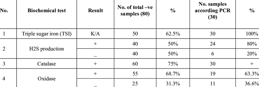
7. Triple Sugar Iron (TSI) Agar Test
Based on their capacity to ferment glucose, lactose, and sucrose as well as generate hydrogen sulfide, bacteria are distinguished using the TSI test.
Method:
- Through stabbing the butt and streaking the slant, the bacteria is introduced into the TSI agar slant.
- Color changes and gas generation are used to interpret the data after incubation.
- Typically, B. cepacia exhibits an acid butt (K/A) and an alkaline slant without producing hydrogen sulfide.
8. Gelatin Hydrolysis Test
The ability of the organism to create gelatinase, which dissolves gelatin, is evaluated in this assay. Gelatinase is not present in B. cepacia.
Method:
- After being injected into gelatin stab tubes, the bacteria is cultured.
- Positive outcome is indicated by the gelatin’s liquefaction.
9. Lysine Decarboxylase Test
The enzyme that converts lysine to cadaverine is detected by the lysine decarboxylase test, which is a crucial characteristic for B. cepacia.
Method:
- The bacteria are introduced into a lysine-containing media.
- A successful outcome is shown by a purple tint following incubation. Generally, B. cepacia is positive for lysine decarboxylase.
Conclusion
Burkholderia cepacia can only be correctly identified through biochemical testing. An extensive profile that aids in differentiating B. cepacia from other bacteria is provided by the combination of tests for oxidase, catalase, indole, MR-VP, citrate utilization, urease, TSI, gelatin hydrolysis, and lysine decarboxylase. Precise detection is essential for efficient handling and therapy of infections, especially in susceptible patient groups like those suffering from cystic fibrosis.
Frequently Asked Question(FAQ)
How do you test for Burkholderia cepacia complex?
In order to determine whether Burkholderia cepacia Complex is present in the sample, the USP <60> Test first enriches it in the proper enrichment broth before streaking it onto selective media.
What is the old name for Burkholderia cepacia?
Pseudomonas cepacia
What color is Burkholderia cepacia?
greenish- brown with a yellowish halo or white with a yellowish-pink halo
Related Article