
The brightfield microscope, also referred to as the compound light microscope, is a pivotal instrument used in various biological and medical research applications. Its functionality is based on the transmission of light through a specimen, with the resulting image being magnified by a series of lenses. Profound knowledge of its intricate components and their respective functions is imperative for proficiently utilizing the microscope to observe and analyze minute microscopic specimens.
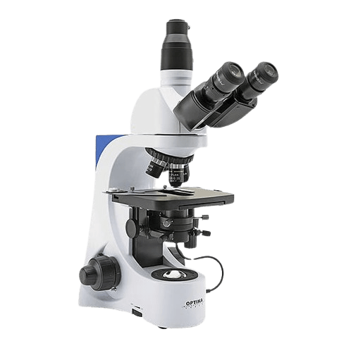
Table of Contents
Definition of Brightfield Microscope
A brightfield microscope, also known as a compound light microscope, is a type of optical microscope widely used in laboratories and educational settings for magnifying and observing specimens. This microscope employs visible light and a series of precisely engineered lenses to produce magnified images of microscopic objects. The term “brightfield” refers to the method’s use of illumination from below, causing specimens to appear dark against a bright background, which enhances contrast and visibility. It is versatile and can be used for various applications, including biological and medical research, as well as educational purposes.
Principle of Brightfield Microscope
The principle of a brightfield microscope involves passing light through a specimen, which is then magnified by a series of lenses. The light source is usually located at the base of the microscope and it passes through the specimen. The image is then magnified by the objective lenses and further enlarged by the eyepiece lenses, allowing for detailed visualization of the specimen.
Key Steps in the Principle:
Illumination
Light is emitted from a built-in light source, typically a halogen or LED bulb. The light is then directed through the condenser, which focuses it onto the specimen.
Transmission
The light passes through the specimen, which may absorb, transmit, or refract the light in varying degrees, creating contrast. Parts of the specimen that absorb more light appear darker, while areas that transmit light appear brighter.
Magnification
The light passing through the specimen is magnified by the objective lens and then further enlarged by the eyepiece (ocular lens) for observation by the viewer.
Resolution
The resolution of a microscope refers to its ability to distinguish two closely spaced points as separate entities, and it depends on the quality of the lenses and the wavelength of light used.
Parts of a Brightfield Microscope
Base
The bottom part of the microscope provides stability and houses the light source.
Light Source
Usually, a built-in illuminator, such as a halogen or LED bulb, that provides the necessary light for viewing the specimen.
Condenser
A lens system located below the stage focuses light onto the specimen. It often includes an adjustable diaphragm to control light intensity and contrast.
Stage
The flat platform where the specimen slide is placed. It often includes clips or mechanical stage controls to hold and move the slide.
Objective Lenses
Multiple lenses with varying magnification powers (e.g., 4x, 10x, 40x, 100x) mounted on a rotating nosepiece. These lenses are closest to the specimen.
Nosepiece
The rotating part that holds the objective lenses, allows the user to switch between different magnification levels.
Body Tube
The tube that connects the eyepiece to the objective lenses. It ensures proper alignment of the optical components.
Frequently Asked Question
What is a brightfield microscope?
A brightfield microscope, also known as a compound light microscope, is an optical microscope that uses visible light and multiple lenses to magnify and view specimens. The specimen appears dark against a bright background.
How does a brightfield microscope work?
The microscope works by transmitting light through a specimen. The light is focused by the condenser, passes through the specimen, and is then magnified by the objective lenses and the eyepiece lens. This magnified image is viewed through the eyepiece.
What are the different magnifications available in a brightfield microscope?
Brightfield microscopes typically have multiple objective lenses with magnifications such as 4x, 10x, 40x, and 100x. Combined with a 10x eyepiece lens, total magnifications of 40x, 100x, 400x, and 1000x can be achieved.
Related Articles