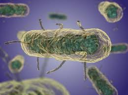
The plague’s causal agent, Yersinia pestis, may be distinguished from other Yersinia species and similar bacteria using a variety of biochemical assays.
These examinations comprise an array of assays designed to evaluate the specific metabolic and enzymatic functions of Yersinia pestis. Important diagnostics include the negative oxidase test and the Gram stain, which identify it as Gram-negative coccobacilli. Other assays that provide unique insights into its metabolic profile include the Triple Sugar Iron (TSI) agar test, Voges-Proskauer, methyl red, urease, catalase, and indole tests. Additionally, the accuracy of identification is improved by motility tests, growth characteristics on selective medium such as CIN agar, and molecular techniques like as PCR.
Table of Contents

Here are some of the key biochemical tests and their expected results for Yersinia pestis
1. Gram Stain in Biochemical Test of Yersinia pestis

The Gram stain is an essential first test in the biochemical testing process for Yersinia pestis. Gram-negative coccobacilli, or rod-shaped bacteria with a somewhat rounded appearance, are the physical manifestation of Yersinia pestis. Its bipolar staining, which presents a “safety pin” look when the extremities of the bacterium stain more brightly than the center, is a distinguishing characteristic. This is especially apparent when using specific stains like Wayson or Giemsa. The early detection and distinction of Yersinia pestis from other Gram-negative bacteria is facilitated by its distinctive staining pattern.
2. Oxidase Test in Biochemical Test of Yersinia pestis
An essential biochemical test for identifying Yersinia pestis is the oxidase test. The cytochrome c oxidase enzyme’s existence in the bacterial cells is ascertained by this assay. The oxidase test results for Yersinia pestis are negative, meaning that the bacteria does not generate the cytochrome c oxidase enzyme. The bacteria are exposed to a reagent, such as tetramethyl-p-phenylenediamine, during the test. Within a few seconds, the reagent will turn dark blue or purple if the bacteria generate cytochrome c oxidase. On the other hand, Yersinia pestis exhibits no color change, indicating that it is oxidase-negative. Yersinia pestis can be distinguished from other oxidase-positive bacteria thanks to this negative finding.
3. Catalase Test in Biochemical Test of Yersinia pestis
One crucial biochemical test for identifying Yersinia pestis is the catalase test. The catalase enzyme, which converts hydrogen peroxide into water and oxygen, is found with this assay. Since Yersinia pestis generates the enzyme catalase, it is catalase positive. A little quantity of the bacterial isolate and hydrogen peroxide are combined for the test. If the bacteria secrete catalase, oxygen bubbles burst quickly, resulting in an observable effervescence. This positive response helps distinguish Yersinia pestis from species that lack catalase and indicates the existence of catalase.
4. Urease Test in Biochemical Test of Yersinia pestis
One important biochemical test for identifying Yersinia pestis is the urease test. This test evaluates the bacterium’s capacity to produce the enzyme urease, which hydrolyzes urea into ammonia and carbon dioxide. The urease test results for Yersinia pestis are negative, meaning that the bacteria does not make urease. The bacterial isolate is injected into a medium containing urea and a pH indicator during the test. The presence of urease causes urea to be hydrolyzed, which causes an alkaline shift that modifies the pH indicator’s hue. Yersinia pestis, on the other hand, exhibits no color change, indicating that it is urease-negative. This negative outcome aids in distinguishing Yersinia pestis from other bacteria that are urease-positive.
5. Indole Test in Biochemical Test of Yersinia pestis
An important biochemical test for Yersinia pestis identification is the indole test. This test assesses the bacterium’s capacity to use the enzyme tryptophanase to convert the amino acid tryptophan into indole. The indole test findings for Yersinia pestis are negative, meaning that the bacteria is unable to convert tryptophan into indole because it is unable to make tryptophanase. Kovac’s reagent is introduced after the bacterial isolate has been cultivated in a tryptophan-containing medium for the test. A good response would cause the medium’s top to become red or pink, signifying the presence of indole. Yersinia pestis, on the other hand, exhibits no color change, indicating that it is indole-negative.
6. Methyl Red Test in Biochemical Test of Yersinia pestis
One important biochemical test for identifying Yersinia pestis is the Methyl Red (MR) test. This test evaluates the bacterium’s capacity for mixed acid fermentation, which results in the production of stable acidic end products from the metabolism of glucose. The Methyl Red test for Yersinia pestis delivers a positive result, meaning that the bacteria generates enough acid to bring the medium’s pH down. The bacterial isolate is cultured in a buffered glucose broth throughout the test. Methyl red, a pH indicator, is added after incubation. Yellow denotes a negative outcome (neutral to slightly acidic environment), whereas persistent red suggests a good result (low pH, acidic environment).
7. Voges-Proskauer Test in Biochemical Test of Yersinia pestis
One crucial biochemical test for identifying Yersinia pestis is the Voges-Proskauer (VP) test. This test establishes if the bacteria can ferment glucose using the butanediol route to create acetoin, a neutral end product. The VP test results for Yersinia pestis are negative, meaning that the bacteria does not make acetoin. The test involves culturing the bacterial isolate on a glucose-rich medium, followed by the addition of potassium hydroxide and alpha-naphthol. The presence of acetoin is indicated by a positive result, which is shown by the development of a red or pink hue. On the other hand, Yersinia pestis exhibits no color change, indicating that it is VP-negative. This negative outcome aids in distinguishing Yersinia pestis from VP-positive bacteria that digest food to create acetoin.
8. Citrate Utilization Test in Biochemical Test of Yersinia pestis
One important biochemical test for identifying Yersinia pestis is the citrate utilization test. This test assesses the bacterium’s capacity to grow only on citrate as its carbon source. The citrate utilization test for Yersinia pestis produces a negative result, meaning that the bacteria is unable to use citrate. Simmon’s citrate agar, which has citrate as the only carbon source and bromothymol blue as the pH indicator, is infected with the bacterial isolate during the test. The medium becomes blue when the bacteria is able to consume citrate because of the alkaline byproducts that are created. Yersinia pestis, on the other hand, does not exhibit any color change and its medium stays green, indicating that it is unable to use citrate.
9. Triple Sugar Iron (TSI) Agar Test in Biochemical Test of Yersinia pestis
In order to identify Yersinia pestis biochemically, the Triple Sugar Iron (TSI) agar test is crucial. This test yields important details on the bacterium’s capacity to produce gas and hydrogen sulfide (H2S), as well as to ferment carbohydrates such as glucose, lactose, and sucrose.
Several observations are obtained in the Yersinia pestis TSI agar test. First off, the agar’s slant usually stays alkaline and turns red, signifying that Yersinia pestis is neither a lactose or sucrose fermenter. On the other hand, the agar’s butt becomes yellow and acidic, indicating that glucose is fermented and acid is produced. It is easier to differentiate Yersinia pestis from other bacteria that can ferment a wider variety of sugars thanks to this variation in fermentation patterns.
10. Motility Test in Biochemical Test of Yersinia pestis
A noteworthy evaluation of the bacterium’s motility in the biochemical testing of Yersinia pestis is the motility test. In particular, Yersinia pestis has a distinct motility pattern that varies with temperature. Yersinia pestis exhibits movement at temperatures of around 25°C, which are similar to those in the flea vector. This motility is frequently observed as a spreading growth pattern on agar plates. Nonetheless, Yersinia pestis is usually non-motile at 37°C, which is crucial to its pathogenicity in humans.
This unique motility behavior helps identify and comprehend the life cycle and pathogenicity of Yersinia pestis. For survival and transmission dynamics, the capacity to transition between motile and non-motile phases in response to environmental stimuli is essential.
11. Growth Characteristics in Biochemical Test of Yersinia pestis
Understanding Yersinia pestis’s development characteristics is essential for biochemical tests in order to distinguish it from other bacteria and identify it. The ideal temperature range for Yersinia pestis to flourish is between 28 to 30°C, which is similar to what natural hosts like fleas and rodents experience. Upon cultivation on conventional media like blood agar and MacConkey agar, Yersinia pestis displays unique growth characteristics. It is noteworthy because it does not ferment lactose, a property that is seen on media that include lactose, such as MacConkey agar, where it manifests as colonies that do not digest lactose.
Furthermore, as demonstrated by assays such as Triple Sugar Iron (TSI) agar, which exhibits an acidic yet alkaline slant, Yersinia pestis exhibits glucose fermentation with acid generation.
12. Selective Media in Biochemical Test of Yersinia pestis
In order to identify and analyze Yersinia pestis biochemically, selective media are essential. A frequently utilized selective medium is CIN agar, which stands for Cetofinosan-Irgasan-Novobiocin agar. The inhibitors in CIN agar let Yersinia pestis to grow unhindered while selectively inhibiting the development of other bacteria. The medium’s cefsulodin, irgasan, and novobiocin components prevent the development of many gram-negative and gram-positive bacteria, which favors Yersinia pestis by establishing a selective environment.
Upon inoculation into CIN agar, Yersinia pestis produces distinct colonies that facilitate identification. These colonies may have a colorless to pink-red appearance because Yersinia pestis is not fermenting lactose.
13. Molecular Methods in Biochemical Test of Yersinia pestis
The biochemical testing and conclusive identification of Yersinia pestis depend heavily on molecular techniques. Polymerase Chain Reaction (PCR) is a particularly important technique among them. By amplifying certain genetic sequences linked to the bacterium, such as the plagene (encoding the plasminogen activator) and the caf1 gene (encoding the F1 capsule antigen), PCR enables the quick and precise identification of Yersinia pestis. The presence of these genes, which are extremely unique to Yersinia pestis, verifies the bacterium’s identification. High sensitivity and specificity can be achieved by using molecular methods such as PCR, which make it possible to find even minute quantities of bacterial DNA in environmental or clinical samples. Furthermore, compared to conventional culture-based procedures, these technologies can produce data considerably more quickly, allowing for quicker identification
summary of Biochemical Test of Yersinia pestis
The biochemical test of Yersinia pestis is crucial for its identification. This comprehensive biochemical test of Yersinia pestis includes the Gram stain, showing Gram-negative coccobacilli, and the oxidase test, which is negative. Another key biochemical test of Yersinia pestis is the catalase test, which is positive, while the urease and indole tests, both part of the biochemical test of Yersinia pestis, are negative. The methyl red test shows positive results, whereas the Voges-Proskauer test is negative. The citrate utilization test, part of the biochemical test of Yersinia pestis, is negative, and the TSI agar test shows an acidic butt and alkaline slant. The motility test, included in the biochemical test of Yersinia pestis, shows non-motility at 37°C. Additionally, selective media and molecular methods enhance the biochemical test of Yersinia pestis, ensuring accurate identification and diagnosis.
Frequently Asked Question
1. How do you identify Yersinia pestis?
Principal traits of Yersinia pestis: Gram stain structure: Rods with Gram negativity, 0.5 x 1-2 µm Colony morphology: non-lactose fermenter on MAC/EMB; growing at both 25–28°C and 35–37°C; colonies are 1-2 mm, gray-white to slightly yellow, and opaque on BAP after 48 hours.
2. Is Yersinia pestis catalase positive?
Yersinia are bacteria that are oxidase-negative, catalase-positive, whose cells are primarily Gram-negative straight rods.
3. Is Yersinia pestis positive or negative?
Yersinia pestis is a small, Gram-negative coccobacillus, which frequently shows strong bipolar staining.
4. what is Biochemical Test of Yersinia pestis
The biochemical test of Yersinia pestis refers to a series of laboratory assays used to identify and differentiate this bacterium from other organisms based on its unique biochemical and metabolic characteristics. Yersinia pestis, the causative agent of plague, can be distinguished through various tests that examine its enzymatic activities, sugar fermentation abilities, and growth patterns.
5. How is Yersinia pestis identified in the lab?
Smears for staining may be prepared in order of likely positive results (i.e., cultures, bubo aspirates, tissue, blood, and sputum specimens). b. Characteristics: Direct microscopic examination of specimens and cultures by Gram stain can provide a rapid presumptive identification. Stained specimens containing Y.
Related Articles

