
Albert Staining is a special staining technique used primarily for the visualization of metachromatic granules (also known as volutin or Babes-Ernst granules) within bacterial cells, particularly in Corynebacterium diphtheriae. These granules are composed of polymetaphosphate and are characteristic of certain bacteria.
Over time, certain stains have been created to help with species identification, morphological differentiation, and even characterization of their unique characteristics. The three most used Albert Staining types are endospore, acid-fast, and gram. Based on their morphologies, each of these stains seeks to identify and describe different types of bacteria.
Albert Staining functions similarly. Its use is intended to detect bacteria that have unique features called metachromatic granules. Neser’s’ stain and Pugh’s stain are two further staining methods that are used to identify granules in bacterial cytoplasmic membranes.
Table of Contents
Albert Staining
Principle
Albert Staining takes advantage of the metachromatic properties of the granules. Metachromatic granules stain a different color compared to the rest of the bacterial cell due to their chemical composition. The staining technique involves the use of specific dyes that bind to the granules, resulting in a distinct color change that allows for their visualization under the microscope.
The goal of the Albert Staining procedure is to find Corynebacterium diphtheriae metachromatic granular masses. Two staining solutions, known as Albert Solution 1 and Albert Solution 2, comprise the Albert stain. Their respective compositions are as follows:
Albert First Solution: Alcohol, glacial acetic acid, malachite green, and toluidine blue
Albert’s second solution: Water containing iodine and potassium iodide
Reagents
The primary stain, Albert Stain I:
- Blue Methylene: 0.15 g
- Green malachite: 0.20 g
- 1.0 ml of glacial acetic acid
- 100 ml of distilled water
Albert Stain II, the emperor:
- 2.0 g of iodine
- 3.0 g of potassium iodide
- 300 ml of distilled water
Procedure
Setting Up the Smear: On a sterile glass slide, spread a thin layer of the bacterial sample.
After letting the stain air dry, heat-fix it by running it over a flame.
Discoloration: Apply Albert Stain I liberally to the smear and let it stand for three to five minutes.
Use distilled water to gently wash the smear.
After applying Albert Stain II liberally, let the smear stand for one to two minutes.
After carefully washing the smear with distilled water, let it air dry.
Observation: Examine the stained smear with the oil immersion objective (100x) in a microscope.
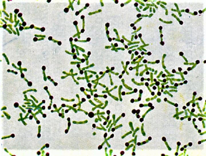
Results
- Granules that are metachromatic will have a dark blue to black appearance.
- Cytoplasm the bacterial cell’s remaining portion will seem green.
Interpretation
- The green bacterial cells include dark blue to black granules, which are indicative of metachromatic granules found in Corynebacterium diphtheriae and related bacteria.
- These granules may be easily identified and differentiated from the rest of the cell due to their unusual color, which makes them useful for studying and diagnosing bacteria that contain them.
Albert Staining is a valuable tool in microbiology for identifying and studying bacteria with metachromatic granules, particularly in clinical and research settings focused on infectious diseases.
Albert Staining is a critical diagnostic tool in microbiology, particularly for identifying Corynebacterium diphtheriae through the visualization of metachromatic granules. The technique’s specificity hinges on the metachromatic properties of these granules, which, upon staining, exhibit a distinctive dark blue to black coloration against the green cytoplasm of the bacterial cells
Frequently Asked Questions(FAQ)
What is Albert Staining?
Albert Staining is a special staining technique used primarily for the visualization of metachromatic granules (also known as volutin or Babes-Ernst granules) within bacterial cells, particularly in Corynebacterium diphtheriae
What is the goal of albert staining procedure?
The goal of the Albert Staining procedure is to find Corynebacterium diphtheriae metachromatic granular masses
What is the result obtained from Albert Staining?
Granules that are metachromatic will have a dark blue to black appearance.
Cytoplasm the bacterial cell’s remaining portion will seem green.
What is Albert Staining I and II solution?
Alcohol, glacial acetic acid, malachite green, and toluidine blue, Water containing iodine and potassium iodide
Related Article