
Fungal diseases can be challenging to diagnose due to their often nonspecific symptoms and the wide range of fungi that can cause infections. Accurate diagnosis is crucial for effective treatment. Here’s an overview of clinical, conventional, and molecular approaches for diagnosing fungal diseases.
Table of Contents
Different Approaches to Fungal Disease Diagnosis
Fungal diseases can be difficult due to their ambiguous symptoms and the range of fungi that cause infection. Accurate and prompt diagnosis is essential for successful treatment. Here is a comprehensive summary of clinical, traditional, and molecular methods for diagnosing fungal illnesses.
1. Clinical Approaches:
Clinical Diagnosis:
Patient’s History:
Symptoms: Collect specific information about the symptoms (such as fever, cough, and skin lesions) and their duration.
Inquire about any potential fungal spore exposure, such as recent travel, employment hazards, hobbies (for example, gardening), and interaction with animals.
Risk Factors: Assess for risk factors such as immunosuppression (e.g., HIV/AIDS, chemotherapy), diabetes, recent operations, or corticosteroid use.
Physical Exam:
Visual Inspection: Check the skin, nails, and mucous membranes for symptoms of fungal infection, such as rashes, discolouration, or sores.
Systemic Symptoms: Look for indicators of infection, such as fever, lymphadenopathy, and respiratory distress.
Perform particular physical examinations based on the probable fungal infection (for example, check for neck stiffness in cryptococcal meningitis).
Clinical Assessment:
- Differential Diagnosis: Consider other possible causes of symptoms (e.g., bacterial or viral infections) and use clinical judgment to narrow it down to a fungal infection.
- If clinical suspicion is high, initiate empirical antifungal medication while waiting for confirming tests.
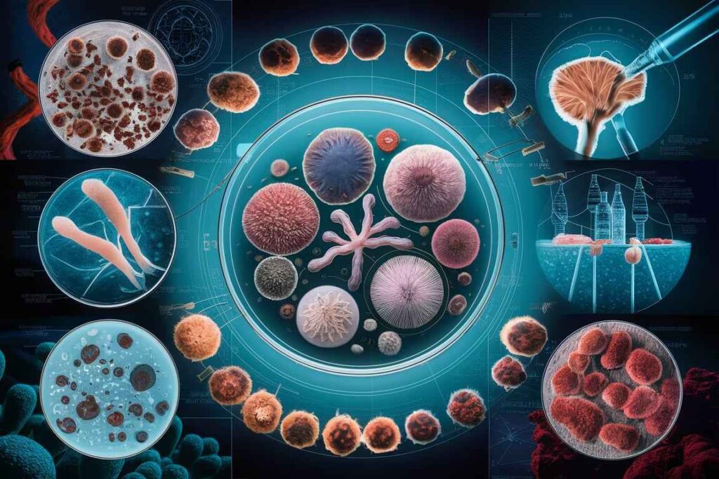
2. Conventional approaches
Microscopy:
1. Direct microscopy:
- Staining: To visualize fungal elements in clinical samples, use stains such as potassium hydroxide (KOH) preparation, Gram stain, India ink (for Cryptococcus), and Calcofluor white.
- Fluorescence microscopy: Calcofluor white stain binds to fungal cell walls and fluoresces under UV light, improving visibility.
2. Histopathology:
- Tissue biopsies involve the examination of tissue sections stained with Periodic Acid-Schiff (PAS) or Gomori Methenamine Silver (GMS) to detect fungi in tissue samples.
- Morphology identifies distinctive fungal structures, such as hyphae and spores, to aid in diagnosis.
3. Molecular Methods:
Polymerase Chain Reaction (PCR).
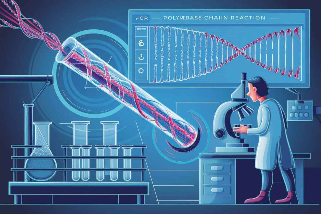
1. Conventional PCR:
- DNA amplification: Increases the amount of fungal DNA in clinical specimens, allowing for more quick and specific diagnosis.
- Primers: Use specialized primers to target conserved fungal genes (e.g., ITS regions, 18S rRNA).
2. Real-time PCR (qPCR)
- Quantification: Provides quantitative information on fungal burden.
- Rapid Diagnosis: A faster turnaround time than traditional culture methods.
Nucleic Acid Sequence:
Sanger Sequencing:
- Identification involves sequencing certain genetic areas to identify fungal species.
- Target regions include the internal transcribed spacer (ITS) regions, the 18S rRNA gene, and the large subunit rRNA gene’s D1/D2 regions.
Next Generation Sequencing (NGS):
- Comprehensive analysis enables the detection and identification of several fungal infections in a single run.
- Metagenomics is useful for assessing complex clinical samples with heterogeneous fungal communities.
Molecular Typing:
- Techniques: Restriction Fragment Length Polymorphism (RFLP), Multilocus Sequence Typing (MLST), Random Amplified Polymorphic DNA (RAPD).
- Epidemiological Studies: Used for strain typing and tracking the source of fungal outbreaks.
Mass Spectrometry
MALDI-TOF MS:
- Protein Fingerprints: Identifies fungi by analyzing unique protein patterns.
- Speed: Provides rapid and accurate identification.
Advantages of Molecular Methods:
- Speed: Faster results compared to conventional methods.
- Sensitivity and Specificity: Higher sensitivity and specificity, especially for detecting low fungal burden and mixed infections.
- Comprehensive: Ability to detect and identify a broad range of fungal pathogens.
Limitations of Molecular Methods:
- Cost: Higher costs associated with equipment and reagents.
- Expertise: Requires specialized training and expertise.
- Contamination Risk: High sensitivity can lead to contamination and false-positive results.
Frequently Asked Question
Define Fungal Disease.
Fungal Disease , or mycoses, are infections caused by fungi, a varied group of microorganisms that includes yeasts, molds, and mushrooms. Fungal infections can affect the skin, nails, respiratory system, and internal organs. Fungal Disease infections can vary from superficial, affecting the skin or mucous membranes, to systemic, affecting internal organs and possibly fatal, particularly in immunocompromised people.
What are advantages of Molecular Methods?
The advantages of Molecular Methods are
1. Speed: Faster results compared to conventional methods.
2. Sensitivity and Specificity: Higher sensitivity and specificity, especially for detecting low fungal burden and mixed infections.
3. Comprehensive: Ability to detect and identify a broad range of fungal pathogens
Related Article
Rickettsia diseases: Pathogenesis, Typhus fever group, Spotted fever group